In the last years, I and multiple other VASO users have encountered many occasions of voxels that show negative CBV change (positive VASO signal change) while the BOLD suggests that the activation should be positive. In this blog post, I want to list potential sources of this surprising effect.
1.) Inflow
VASO is an inversion-recovery sequence, and is thus very similar to an ASL-CBF sequence. Similarly, like in ASL, VASO can therefore have contaminations of inflowing fresh (uninverted) blood. E.g. in areas of very short arterial arrival times (e.g. large arteries), fresh blood can flow into the imaging region. The voxels that are effected by this inflow, are usually very bright in the raw VASO signal and can be easily identified. During activity increase, more of this fresh blood flows into the imaging regions and functionally increases the resulting VASO signal dependent on the CBF increase. Since VASO is a negative CBV contrast, this inflow effect counteracts the CBV related VASO signal change and canmask negative signal change.

This effect of inflow-related negative voxels is easily identified and hence can be accounted for by excluding these voxels from the analysis. The effect can be removed form the acquisition side by choosing shorter inversion times (at the cost of SNR). Since this effect is usually only happening in pial vessels, it can be ignored for analyses pipelines that focus on GM voxels only.
2.) Venous constriction
The skull can be considered as a container with a fixed volume and cannot be expanded. As soon as one compartment increases its volume, another compartment needs to reduce its volume accordingly. Until now its it still not really established how this mechanism works and how the substantial CBV increase during increased brain activity is accounted for. Dependent on the task duration, activation region size, and activation strengths, CBV increase seems to be jointly compensated by GM-volume decrease, CSF-volume decrease and decrease of venous blood volume.
The last option, the decrease of venous blood volume, can thus also be one possible reason for negative VASO signal change and has been described here.


In my experience, this effect is more common than inflow effects and it is mostly happening in the upper layers and above the cortex.
I found that these voxels are most easily identified by mapping the correlation of the VASO and the BOLD fluctuation. E.g. with the LAYNII program LN_CORREL2FILES.

3.) Errors in the BOLD correction
For almost any VASO imaging readout, there is a finite echo time and a BOLD contamination needs to be corrected for. In my experience, this is done with simultaneous acquisition of images with and without blood nulling. The BOLD-correction the consist of a simple division e.g. with the formula VASO_n = (Nulled_n) / (0.5×BOLD_n-1 + 0.5×BOLD_n+1). So, the BOLD of neighboring time points is interpolated to estimate the hypothetical BOLD contamination at the time point, when the VASO image was acquired. Errors in tis interpolation can result in an insufficient BOLD correction and result in negative VASO voxels.

4.) Noise level
Another source of negative VASO voxels can also be noise level. VASO has about 50% of the CNR compared to BOLD, so it the probability is higher that positively activated voxels show negative VASO voxels just because of noise.


5.) CSF-volume redistribution
The T1-sensitivity in VASO makes it not only sensitive to changes in CBV, but it is actually also believed to be sensitive to volume redistribution of any other T1-compartments too. As such, Piechnik at al., Seong-Gi Kim, the Constable group and Manus Donahue proposed that there might be dynamic change of CSF that can contaminate the VASO contrast. In SS-SI VASO this effect can be accounted for with a balanced combination of TRs and flip angles, as such that CSF and GM have the same baseline signal. In this sequence setup, the VASO signal reflects both: (a) the CBV increase that is compensated with extravascular parenchymal CBV and (b) the CBV increase that is compensated with CSF volume decrease.

With this in mind, it shows an additional channel of potential VASO signal increase (negative VASO voxels): CSF volume reduction that are accompanied the GM volume increase.
Across multiple examples of strong tasks, I never found clear evidence for this to happen. A few examples are shown here. The only exception are global tasks, such as respiration challenges.

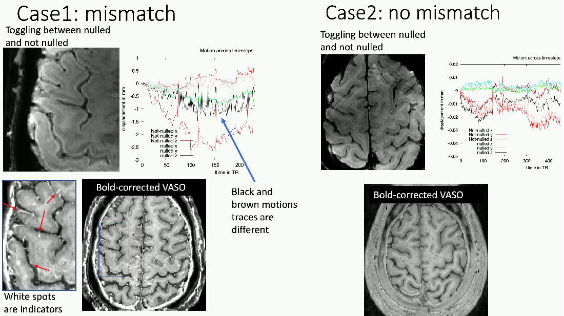
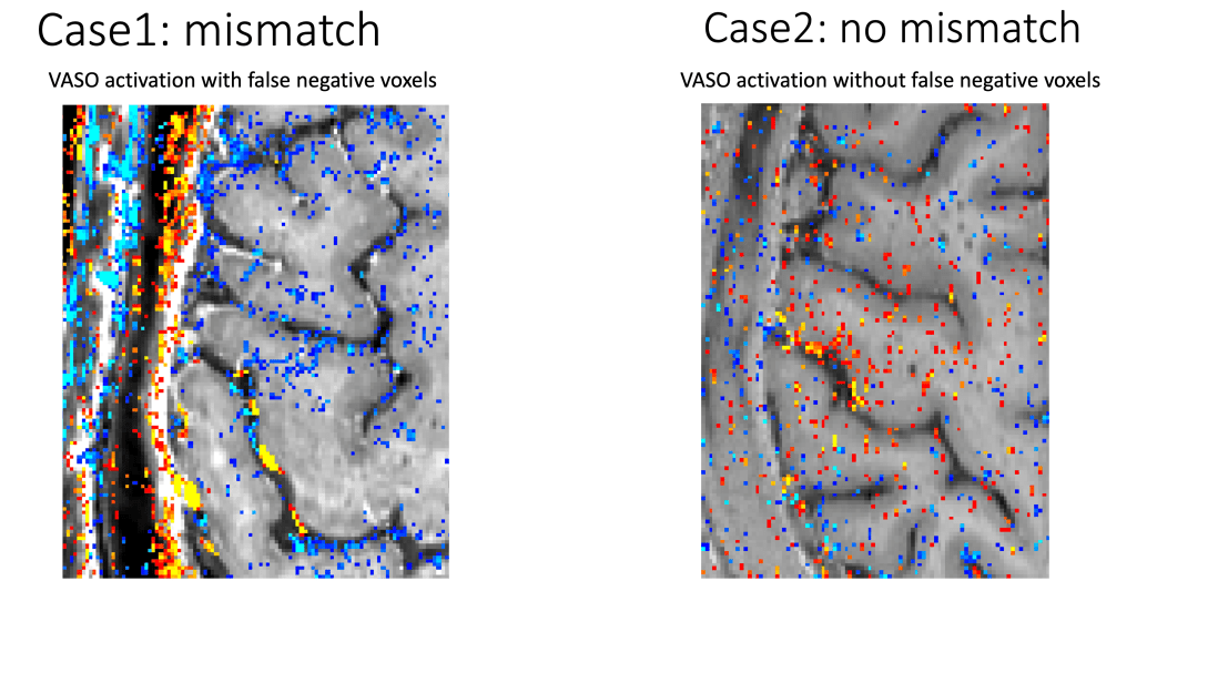
5.) locally missmatched BOLD correction
When the alignment of BOLD and VASO images are not perfectly accurate with respect to each other, it can happen that the BOLD contamination is underestimated in some voxels and overcorrected in other voxels. The latter can result in erroneously negative CBV estimations.