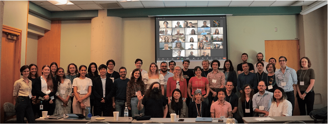
The combination of ultra-high field (7 Tesla and above) imaging with increasingly sophisticated data analysis tools has led to a surge of research using functional MRI acquisitions to examine the behavior of individual cortical layers of the brain. This course will focus on teaching the acquisition and analysis tools needed to contribute to this research.
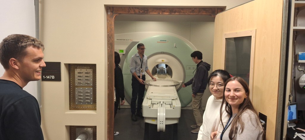
Content: four days of hands-on training on laminar fMRI day 1: introduction to laminar fMRI & basic data acquisition day 2: data preprocessing and analysis day 3: interpretation and modeling day 4: advanced applications and future directions
Authors: Kenny (Chung) Kan1, Rüdiger Strinberg2, and Renzo (Laurentius) Huber3
Functional MRI Facility (FMRIF), NIMH, NIH, USA
German Center for Neurodegenerative Diseases (DZNE), Germany
Martino’s Center for Biomiedical imaging, Mass general Brigham, Harvard Medical School
What happens with the EPI sequence when I increase the TE? Which bandwidth should I use? What do I need to change to reduce PNS? There are many common misconceptions in basic EPI protocols. This blog post discusses the most common ones.
This post is a a continuation of highlighting basic acquisition features of EPI. See posts in GRAPPA, Regularization, Ghost correction, and 3rd order shim here.
This post summarized the layer-fMRI abstracts form major relevant conferences 2024, including ISMRM, OHBM. This is following the posts of previous years 2024, 2023, 2022, 2021, 2020, 2019. Edit suggestions are welcome (renzohuber@gmail.com)
This hackathon project is part of the series, Hack your Scanner, following contributions of previous years. 2022 VASO mosaic, 2021 visual exporting scanner data with QR Modem, 2020 viewing data with ASCII art on MARS with LN_INFO. This year is about hacking your RF-coil.
This post summarized the layer-fMRI abstracts form major relevant conferences 2024, including ISMRM, OHBM. This is following the posts of previous years 2023, 2022, 2021, 2020, 2019.
Functional magnetic resonance imaging (fMRI) today is a common method to study the human brain. The popularity of fMRI can be explained by its versatility and accessibility to study the structure-function relationship in living humans, which has historically been challenging or impossible. One area of constant progress and excitement since the early days in fMRI is the advances in hardware, MR sequences, and software which allowed researchers to image the fine details of the human brain non-invasively. fMRI is currently capable of reaching sub-millimeter resolution, effectively transforming the MRI scanner from a macroscope into a mesoscope. While 0.8 mm isotropic voxel resolution (~0.5 μl) has become a part of the daily routine of a high resolution fMRI researcher at 7 Tesla scanners, as opposed to e.g. 3 mm isotropic voxels in conventional fMRI studies conducted at 3 T (Bollmann et al. 2020), recent developments have breached 0.37 mm isotropic resolution (~0.05 μl) (Feinberg et al. 2023). This 10 fold reduction in voxel volume promises an exciting future to observe the fine mesoscopic details such as cortical layers, columns, and vessels during changes in human brain function.
On Oct 13th 2023, Nicolas Boulant presented an intriguing source of MRI image artifacts at the CMRR high field meeting in Minnesota. He suggested that the 3rd-order shim can result in amplified gradient trajectory imperfections. In low bandwidth FLASH, this can manifest as faint ghosts in the read direction shifted by a few pixels. In EPI, on the other hand, these trajectory errors can result in fuzzy ripples (low spatial frequency ghosts and shadings, not edge ghosts).
In a recent meta analysis of all openly available layer-fMRI datasets, I had found had that the fuzzy ripples are one of the main limits of high-quality layer-fMRI acquisition (see here) across vendors. So, I was curious whether the 3rd order shim might be partly related to this. In this blog post, I am describing my attempts to reproduce Nicola’s results and investigate the effect of the 3rd order shim on layer-fMRI protocols. I find that disconnecting the 3rd order shim can result in significantly better data quality. However, this finding is only visible for specific echo-spacings, which are either in the ‘forbidden frequencies’ or which have side bands in the forbidden frequencies.
This post does not imply that previous research was conducted sub-optimally. Since, it is common practice to optimize the EPI echo spacing in the piloting stage of each study, the frequencies with these artifacts are usually avoided anyway. Here, we confirm that this is a good practice.
On February 8th 2024, we will host the 8th virtual layer-fMRI dinner. As usual, it’s free, just follow the link below.
The 6th layer-fMRI dinner will focus the relationship of layer-fMRI academia and industry. Layer-fMRI basic research and industry have different goals, yet they help and facilitate one another. Without each other, each of our lives would be harder.
Authors: Lasse Knudsen, Luca Vizioli, Federico De Martino, Lonike Faes, Dan Handwerker, Renzo Huber
This post describes the usage, capabilities and challenges of NORDIC PCA denoising on VASO data. A video presentation of this project can be found here: https://youtu.be/bbGKMTWVrJY.
This is the second blog post about graphic representations of cynical metaphors. The first post on graphical metaphors was about finding best layer-fMRI sequence and can be found here. This one is about how layer-fMRI fits into the landscape of other disciplines.
On Dec 7th 2022, we will host the 5th virtual layer-fMRI dinner.
The board of the layer-fMRI dinner group has invited the following speakers to initiate discussions on the theme: Layer-fMRI signal origin: From neurons to vessels to BOLD.
Serge Dumoulin (NINS; Utrecht University): How population receptive field properties change across cortical laminae: from vision to cognition.
Anna Devor (Boston University): Layer-resolved imaging of resting CMRO2 in awake mice with phosphorescent O2 probes.
Lars Muckli (University of Glasgow): From man to non-human primates and mouse: Two top-down streams in different layers of retinotopic visual cortex (the ground truth).
Moderated by Luca Vizioli and Tyler Morgan
The entire event will last for about 90 min (including discussion).
The meeting will be recorded and published on Youtube and embedded on this website by Dec 8, 2022.
Everyone is welcome. No registration required. Zoom link:
Serge Dumoulin How population receptive field properties change across cortical laminae: from vision to cognition.
A key advantage brought by ultra-high field MRI at 7 Tesla and more is the possibility to increase the spatial resolution at which data is acquired, with little reduction in image quality. This opens a new set of opportunities for cognitive neuroscience, for example to probe how signals vary across cortical thickness and laminae. Here, I present recent work on computational modelling of population receptive field properties. I will discuss how they vary across cortical laminae, how these properties are influenced by attention and extend these protocols from primary visual cortex towards numerical cognition in association cortex. I will also discuss some of the limitations and the potential of laminar imaging in human cortex.
Anna Devor Layer-resolved imaging of resting CMRO2 in awake mice with phosphorescent O2 probes.
The cerebral cortex is organized in cortical layers that differ in their cellular density, composition, and wiring. Cortical laminar architecture is also readily revealed by staining for cytochrome oxidase – the last enzyme in the respiratory electron transport chain located in the inner mitochondrial membrane. It has been hypothesized that a high-density band of cytochrome oxidase in cortical layer IV reflects higher oxygen consumption under baseline (unstimulated) conditions. We tested the above hypothesis using direct measurements of the partial pressure of O2 (pO2) in cortical tissue by means of 2-photon phosphorescence lifetime microscopy (2PLM). We revisited our previously developed method for extraction of the cerebral metabolic rate of O2 (CMRO2) based on 2-photon pO2 measurements around diving arterioles and applied this method to estimate baseline CMRO2 in awake mice across cortical layers. Our results revealed a decrease in baseline CMRO2 from layer I to layer IV. This decrease of CMRO2 with cortical depth was paralleled by an increase in tissue oxygenation. Higher baseline oxygenation and cytochrome density in layer IV may serve as an O2 reserve during surges of neuronal activity or certain metabolically active brain states rather than baseline energy needs.
Lars Muckli From man to non-human primates and mouse: Two top-down streams in different layers of retinotopic visual cortex (the ground truth).
Using laminar fMRI, we identified two top-down processing streams. (1) One stream is for contextualizing visual input based on fast recurrent processing of contextual information and (2) a second top-down projection is used for visual imagery. Both top-down streams are based on non-direct geniculate input to visual cortex. To investigate the ‘ground-truth’ of this top-down processing, collaborators in the Human Brain Project (HBP) conducted parallel experiments in monkeys and mice using microelectrode recording and two-photon calcium imaging. The contextual feedback effects in complex visual fields are fast, dependent on learning and are likely communicated by disinhibition in superficial layers of cortex.
Complex cognitive tasks are difficult to instruct in non-human primates and in rodents, but in humans we can see that top-down feedback processing is used for visual imagery, and also object comparison and navigation.
Vascular Space Occupancy is an fMRI method that is popular for high-resolution layer-fMRI. Currently, the most popular sequence is the one by Rüdiger Stirnberg from the DZNE in Bonn, which is actively being employed at more than 30 sites.
This sequence concomitantly acquires fMRI BOLD and blood volume signals. In the SIEMENS reconstruction pipeline, these signals are mixed together within the same time series, which challenges its user friendliness. Specifically: The “raw” dicom2nii-converted time-series are not BIDS compatible (see https://github.com/bids-standard/bids-specification/issues/1001). The order of odd and even BOLD and VASO image TRs is dependent on the nii-converter. Workarounds with 3D distortion correction, results in interpolation artifacts. Workarounds without MOSAIC decorators result in impracticable large data sizes.
The goal of this Hackathon is to extend the 3D-MOSAIC to solve these constraints. This functor is commonly used to sort images by echo-times, by RF-channels, by magnitude and phase in the SIEMENS reconstruction pipeline into sets of mosaics . However currently, this functor does not yet support the dimensionality of SETs. In this project we seek to include SETs into the capabilities of the functor.
This page collects layer-fMRI abstracts from neuro imaging conferences in 2022. This is following the tradition of layer-fMRI abstracts in the previous years: layer-fMRI abstracts 2019, layer-fMRI abstracts 2020, and layer-fMRI abstracts 2021. Comments and completions are welcome (renzohuber@gmail.com).
On Oct 6th 2021, we aim to host the 4th virtual layer-fMRI dinner.
The board of the layer-fMRI dinner group has invited the following speakers to initiate discussions on the theme: Layer-fMRI signal origin: From neurons to vessels to BOLD.
Amir Shmuel (McGill): The complexity of lamina resolved neuronal activity, and the spatial specificity of BOLD, CBV, arterioles and venules responses: implications for planning and interpreting depth-dependent fMRI.
Jonathan Polimeni (MGH): Biophysical modeling for interpreting fMRI signals and relating them back to neuronal activity: contemplating the “inverse problem”.
Evelyn Lake (Yale): Leveraging simultaneous multi-modal fMRI and wide-field optical imaging to study functional brain networks.
Moderated by Luca Vizioli and Andrew Morgan
Board: Johanna Bergmann, Avery Berman, Saskia Bollmann, Denis Chaimow, Renzo Huber, Nils Nothnagel, René Scheeringa, and Bianca van Kemenade.
The entire event will last for about 90 min (including discussion).
The meeting will be recorded and published on Youtube and embedded on this website by October 7th 2021.
Biophysical modeling for interpreting fMRI signals and relating them back to neuronal activity: contemplating the “inverse problem”.
Abstract:
The ultimate limits of spatial and temporal resolution achievable by fMRI are dictated by neurovascular coupling, the mechanisms of blood flow regulation, and vascular architecture in the brain. While these limits are currently unknown, there is a rapidly growing body of evidence pointing to the ability of fMRI to distinguish site of activation across cerebral cortical depths, which can be used to infer the cortical layer or layers differentially engaged in specific tasks or functional networks. Because all fMRI signals currently in use are based on hemodynamics and hence are influenced by local vasculature, understanding how patterns of neural activity are transformed into the fMRI signals we measure can potentially aid not only in the interpretation of our data but also opens possibilities to better estimate the location (in space and time) and amplitude of the neural response from the fMRI response—to the extent that this transformation is “invertible”.
Motivated by this, the goal of this presentation is to survey recent work towards building biophysical models of the fMRI signals to help with this interpretation, with a focus on models using realistic microvascular networks and dynamics based on optical imaging and microscopy data. These models are built on first principles and are described by meaningful anatomical and physiological parameters. I will present initial results demonstrating how these models can be used to predict well- known differences in the hemodynamic response across stimulus configurations and cortical depths. While these models are complex, and simulations are computationally intensive, they can be also used to help inform simpler “lumped” models that are more practical for routine use, and are applicable to predicting various BOLD and non-BOLD fMRI contrasts.
Another goal of this presentation is to engage the laminar fMRI community and have an open discussion about the strengths and weaknesses of this modeling approach, consider these against other approaches to improve neural specificity in fMRI, and discuss how to combine this framework with advanced acquisitions and analysis methods towards our shared objective to measure neural activity across cortical layers with fMRI.
Video recording of Jonathan Polimeni’s presentationAmir Shmuel (McGill)
The complexity of lamina resolved neuronal activity, and the spatial specificity of BOLD, CBV, arterioles and venules responses: implications for planning and interpreting depth-dependent fMRI
Video recording of Amir Shmuel’s presentationEvelyn Lake (Yale)
Leveraging simultaneous multi-modal fMRI and wide-field optical imaging to study functional brain networks.
Coordinator: Jonathan Polimeni, Renzo Huber, and Luca Vizioli
Training Faculty: Laurentius Huber, Rainer Goebel, Anna Izabella Blazejewska, Luca Vizioli, and Jonathan Polimeni
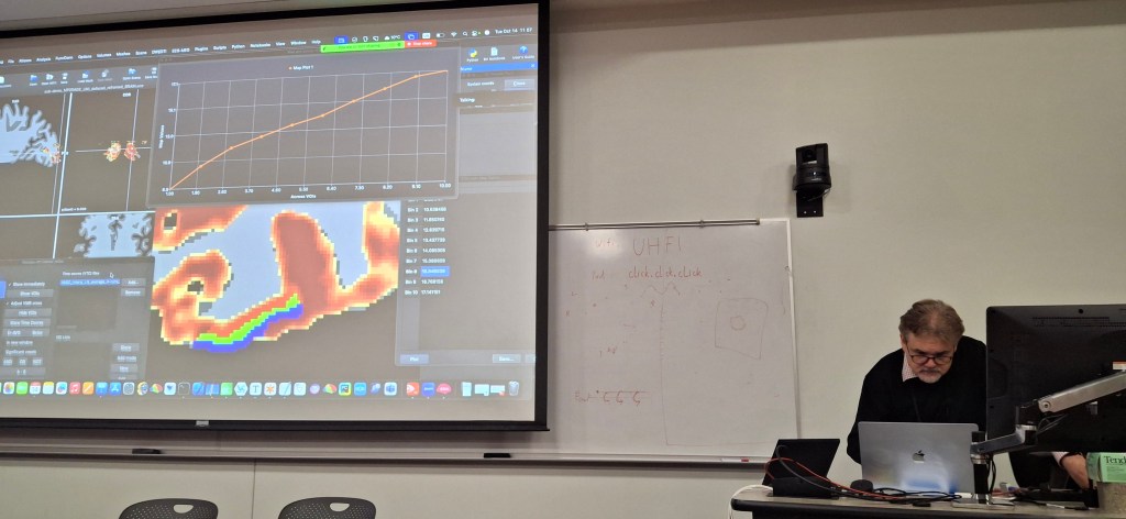
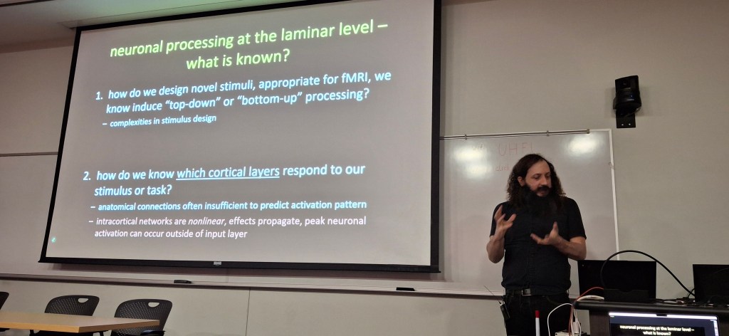
One dataset, many analyses: an overview of the diverse processing approaches in layer-fMRI.
The layer-dinner group would like to invite you to show us your analysis pipeline in a brief presentation at an upcoming “Layer-fMRI dinner” in the Spring of 2022. The analysis of layer-fMRI data is challenging and not straightforwardly doable with standardized streamlines analysis packages. Most layer-fMRI groups have their own dedicated analysis solutions to account for layer-specific challenges. As such, the purpose of this event is:
To illustrate multiple layer analyses of members of the field, and for others to follow.
To highlight challenges of high-res and layer specific analysis.
To stimulate discussion about analysis challenges and solutions.
To give analysis developers a platform to advertise their analysis solutions.
To illustrate differences and similarities of pipelines.
Introduction by Jonathan Polimeni
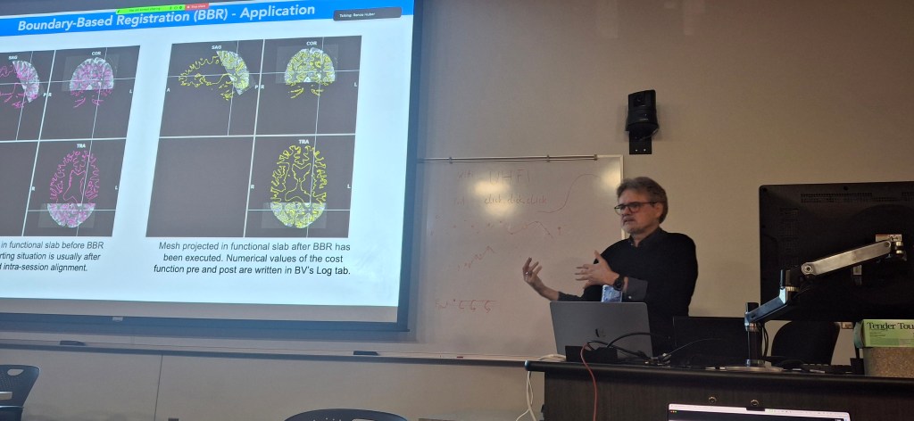
Rainer Goebel: Brain Voyager
Lecture
Hands on
Renzo Huber: LayNii & AFNI
Lecture
Hands on:
Anna Blazejewska: Freesurfer
Lecture
Hand on instructions
For the BrainVoyager hands-on session by Rainer Goebel:
For the FreeSurfer hands-on session by Anna Blazajewska, there are no prior preparations needed. You will get server access. You can access the server with a vnc viewer.
On April 20th 2021, the third virtual layer-fMRI took place. 120 (unique) attendees joined and discussed the connection between layer-fMRI and cognitive models.
This meeting is held as a succession of the first two virtual dinner in May 2020, and Sept 2020:
In this third event, it will be discussed how the layer-fMRI methodologies might be able to inform Cognitive models. The three speakers are researchers that are working to examine cognitive processes whose study is aided by understanding the structure and function of cortical layers. These cognitive processes could include memory, attention, learning, dreaming, language or cortical predictions (plus many, many more!)
Floris de Lange will give an overview of work done by his group to capture laminar fMRI activity changes in the visual cortex for prediction, attention and bottom-up input. André Bastos will present results of laminar LFP recordings and how feed-forward gamma-band and feedback alpha/beta band modulations help to understand cognitive effects including attention, working memory, and prediction processing. Michelle Moerel will talk about how computational models can be combined with laminar fMRI to understand human auditory processing.
Below you find the important links of the virtual event. Embedded videos of the talks, discussions, and a summary of the hot topics are going to be added on the day after the event.
In this second event, it will be discussed how the research field can bridge the gap between layer-dependent activity measures that are obtained with fMRI and electrophysiology, respectively. Kamil Ugurbil will present the perspective of high resolution for human neuroscience, Lucia Melloni will present the perspective of depth-dependent electrophysiological recordings in humans, and Seong-Gi Kim will talk about the combination of both worlds, layer-fMRI and layer-dependent electrophysiological recordings.
Below you find the important links of the the virtual event. Embedded videos of the talks, discussions, and a summary of the hot topics are going to be added on the day after the event.


