In this post I want to describe the guidelines that helped me to find the right spot of primary motor cortex (M1) that has a double-layer pattern during a conventional finger tapping task.
The motor cortex is an excellent model system to debug-layer fMRI methodology for multiple reasons:
- It has a consistent folding pattern across people.
- Its folding pattern is convoluted across one axis only. Hence, it is possible to use thicker slices with higher in-plane resolution.
- With 4mm, its is the thickest part of the cortex compared to all other areas. Hence, layer analysis can be done even with 1.2 mm voxels.
- It has an expected double layer structure, with two separate peaks. The separability of the peaks can be used as a measure of functional specificity.
- It is very close to the RF-receive coils and has high tSNR.
- It is easy to shim.
One tricky part, however, is to find the right location of the double layer feature.
Background


Finding the position
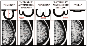
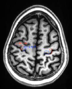
The position within the axial slice is super easy in about 80% of the people. There, however, few people where its not so clear. In those cases, I found it helpful to project the superior frontal sulcus further back. If there is no sulcus in this direction, there is a high likelihood that the selected “omega” structure is actually in the sensory cortex. The superior frontal sulcus is located anterior of the motor cortex. It goes along the anterior-posterior direction and is depicted in green in the examples below.
Below are some examples of how the “omega” structure alone can be misleading. Having the superior frontal sulcus as reference helps. The wrong spots can be identified because they don’t have the superior frontal sulcus anterior to them. Of course, it is helpful to go through many slices to spot the superior frontal sulcus.
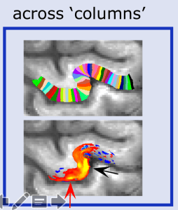
The position in the coronal plane should be placed as far away from the center of the brain as possible.


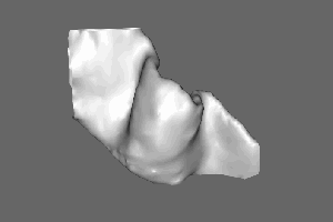
Within the hand knob, you find the double layer response only on the lateral side (M1-4a):


Finding the central sulcus
In some rare instances it can be hard to find the central sulcus. This book chapter

RF considerations for motor cortex
With the 32ch Nova coil at high field strengths (such as 7T), I found that the B1+ field is asymmetric in the area of the motor cortex. Hence, I prefer imaging the right motor cortex during a left-hand motor task. It looks like this is a result of the asymmetric transmit field and corresponding deviations of the optimale flip angle distribution of the 3D-EPI segments.

I found it surprising that the tSNR (in an agar phantom) has a huge heterogeneity across RF channels. Especially the channels close to the motor cortex (11, 15, 0) seem to have a lower tSNR compared to visual areas. However, these channels have the lowest noise coupling between channels, so the tSNR of the combined images is usually not as bad.


