This blog post gives an overview of the scientific network of researchers that are using the VASO (vascular space occupancy) for applications in layer-fMRI. I tried to give an overview of all layer-fMRI VASO papers published so far and provide a map of all layer-fMRI VASO labs around the globe.
Popularity of layer-fMRI VASO across years and countries
The first layer-fMRI VASO studies in humans were presented in 2014/2015. In the five years that followed, VASO became a credible contrast for the emerging field of layer-fMRI.

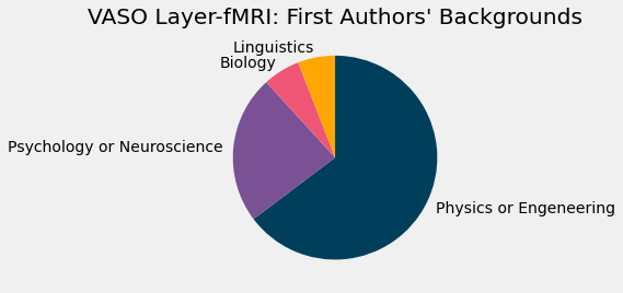
As of today (Jan 2020) there are 31 labs around the world that are using layer-fMRI VASO with 25 published layer-fMRI VASO papers in peer-reviewed journals
The vast majority of layer-fMRI VASO research is happening in Europe, followed by Asia. The higher 7T density in USA is not represented in a correspondingly many layer-fMRI VASO studies. This it might be due to the medical-application driven research funding environment.
Google map to browse interactively: https://layerfmri.page.link/VASO_worldwide (in case of missing sites, suggestions are welcome to layerfmri@gmail.com).
Fifty current users of layer-dependent VASO fMRI
- Ulsan National Institute and Technology, Korea
- Ji-Hyun Kim is using VASO to investigate the laminar patterns of RA and SA columns in S1.

- OHBM 2021 abstract
- Aarhus, Denmark
- Lasse Knudsen and Torben Lund are using layer fMRI VASO at 3T.
- ISMRM 2021
- VA, San Francisco, USA
- An Vu is using layer-dependent VASO and BOLD images and combines them into one ultimate combined measure.
- ISMRM 2021
- NICT, Japan
- Guoxiang Liu is using VASO layer-fMRI with BISEPI at 7T.
- OHBM 2021 talk
- Osaka, Japan.
- Yinghua Yu is using VASO to investigate rhythmic somatosensory prediction.

- OHBM 2021 abstract
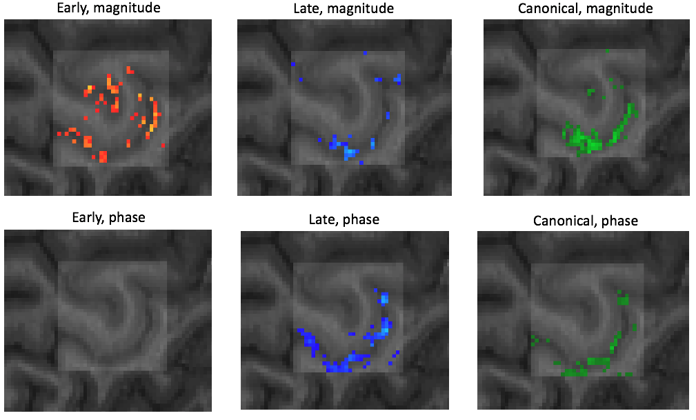
- Max Planck Institute CBS, Leipzig, Germany:
- Daniel Haenelt and Robert Trampel are using VASO (along with GE-BOLD and SE-BOLD) to investigate ocular dominance columns.

This figure kindly provided by Daniel Haenelt and Robert Trampel - Reference: ISMRM 2020
- Daniel Haenelt and Robert Trampel are using VASO (along with GE-BOLD and SE-BOLD) to investigate ocular dominance columns.
- SFIM, NIMH, NIH, Bethesda, USA:
- Yuhui Chai and Peter Bandettini are using VASO as a ground truth method to compare it with the VAPER contrast.
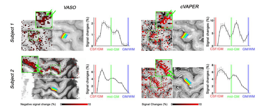
This figure kindly provided by Yuhui Chai - Reference: NeuroImage Paper
- Yuhui Chai and Peter Bandettini are using VASO as a ground truth method to compare it with the VAPER contrast.
- Cardiff University, Cardiff, UK:
- Marcello Venzi, Joseph Whittaker, and Kevin Murphy are using high-resolution VASO to investigate the effect of CSF and veins in superficial voxels vs. parenchyma voxels.
This figure kindly provided by Marcello Venzi - Reference: ISMRM abstract 2019
- Marcello Venzi, Joseph Whittaker, and Kevin Murphy are using high-resolution VASO to investigate the effect of CSF and veins in superficial voxels vs. parenchyma voxels.
- MBIC, Maastricht University, Netherlands:
- Renzo Huber and Benedikt Poser are working on sequence approaches to make layer-fMRI VASO easier applicable.

Whole brain VASO acquisition for easy applicability in neuroscience studies. - Reference: ISMRM abstract 2020, submitted
- Renzo Huber and Benedikt Poser are working on sequence approaches to make layer-fMRI VASO easier applicable.
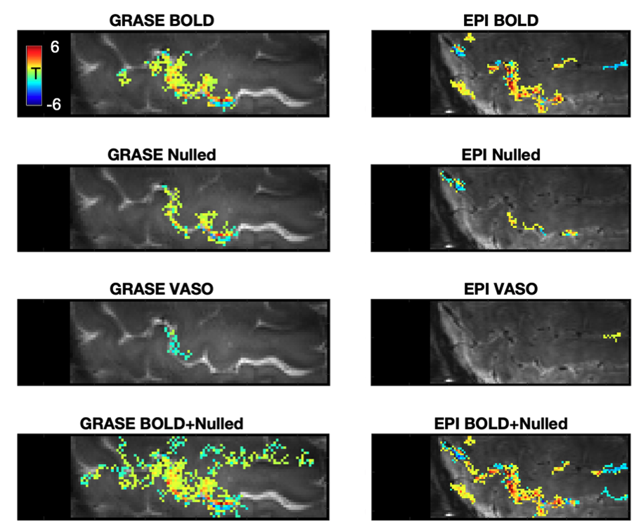
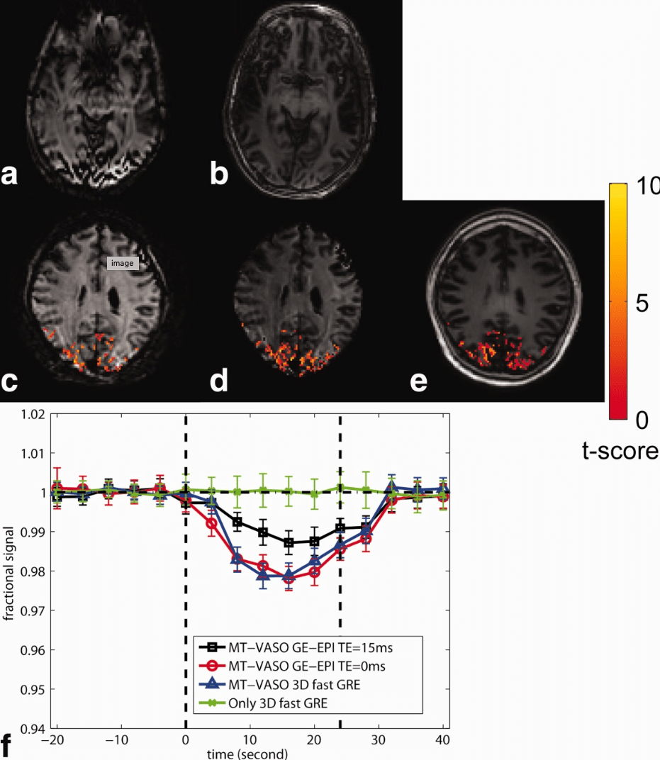
- VA SF, USA:
- Alex Beckett and David Feinberg are using VASO as a ‘gold standard’ to compare it to 3D-GRASE.

-
- This figure is taken from the BioRxiv preprint
-
- Reference: BioArchive Preprint.
- Alex Beckett and David Feinberg are using VASO as a ‘gold standard’ to compare it to 3D-GRASE.
- Spinoza/UMC, Utrecht/Amsterdam, Netherlands:
- Icaro Oliviera, Jorien Siero, and Wietske van der Zwaag are using VASO to investigate the linearity of the hemodynamic response at very high resolutions.

- Reference: Oliviera NeuroImage 2020
- University of Sheffield: Sheffield, UK:
- Aneurin Kennerley is using layer-dependent VASO to validate it against iron-based contrast agent fMRI in rodents.
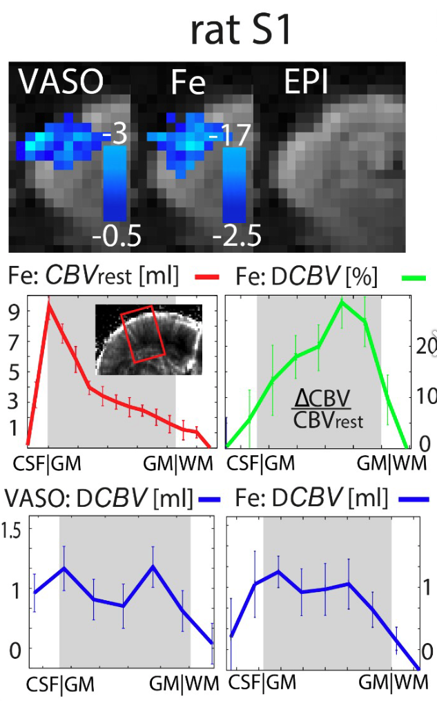
Figure kindly provided by Aneurin Kennerley. - Reference: ISMRM abstract 2017
- Aneurin Kennerley is using layer-dependent VASO to validate it against iron-based contrast agent fMRI in rodents.
- University of York, York, UK
- Aneurin Kennerley and Renzo Huber are working on layer-fMRI VASO to make it doable at 3T.

Figure taken from Kennerley’s submitted ISMRM abstract (2020). - Reference: ISMRM abstract 2020, submitted
- Aneurin Kennerley and Renzo Huber are working on layer-fMRI VASO to make it doable at 3T.
- University of York, UK

- Data kindly provided by Elisa Zamboni and Aneurin Kennerley.
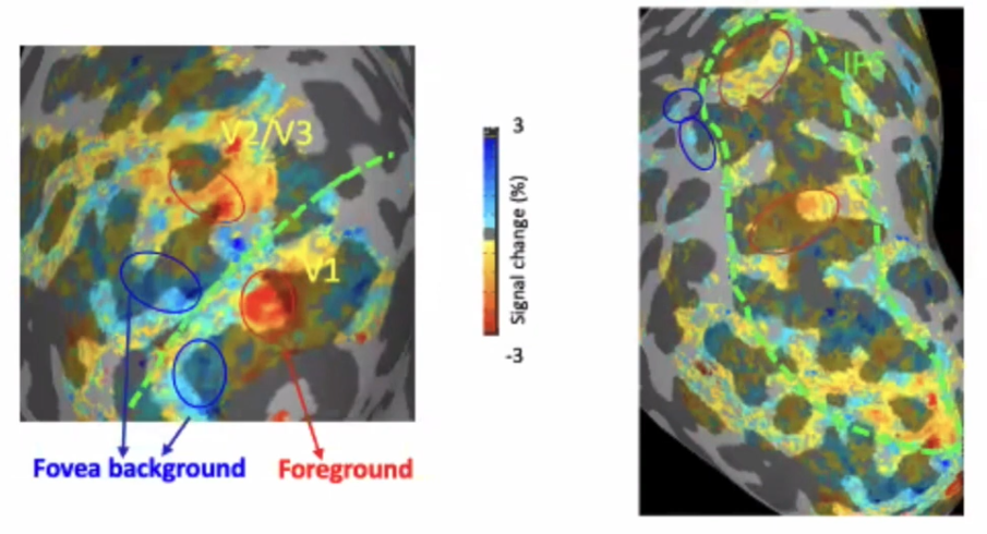
- Lab of Brain and Cognition, NIMH, NIH, Bethesda, USA
- Eli Merriam and Zvi Roth use sub-millimeter VASO to map the visual topography.
This figure is kindly provided by Eli Merriam. - Reference data shown here
- Eli Merriam and Zvi Roth use sub-millimeter VASO to map the visual topography.
- Martinos Center, MGH, Boston, USA:
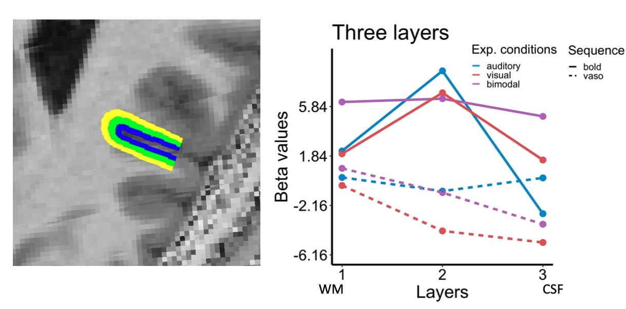
- Saskia Bollmann and Jonathan Polimeni use sub-millimeter VASO to investigate the temporal features of CBV across depth.

This figure is kindly provided by Saskia Bollmann. - Reference data shown here
- Saskia Bollmann and Jonathan Polimeni use sub-millimeter VASO to investigate the temporal features of CBV across depth.
- University of Queensland, Australia:
- Atena Akbari and Markus Barth are investigating the layer-dependent fMRI response of VASO in V1.
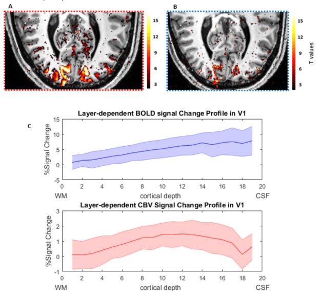
This figure is kindly provided by Atena Akbari. - OHBM abstract 2019
- Atena Akbari and Markus Barth are investigating the layer-dependent fMRI response of VASO in V1.
- University of Glasgow, Glasgow, UK:
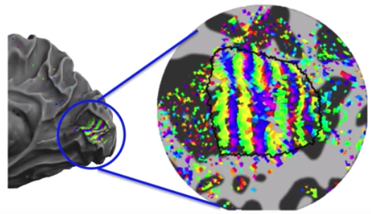
- Nils Nothnagel, Andrew Morgan, and Jozien Goense implemented a 3D-EPI sequence for layer-dependent VASO imaging.
- The first layer-fMRI VASO experiments were conducted early 2019.
-
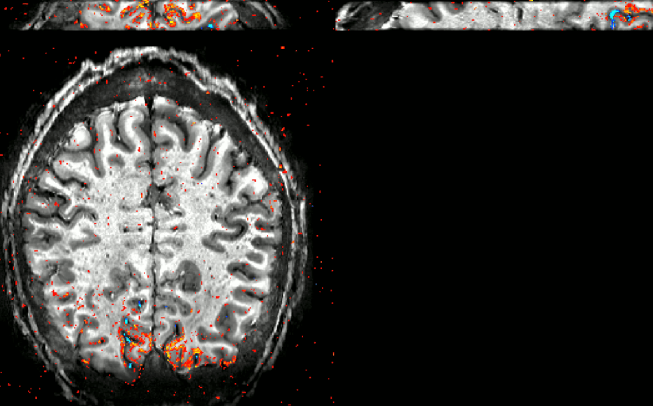
0.6mm VASO during a visual paradigm acquired in Glasgow from Nils Nothnagel and Andrew Morgan
- SKKU, Suwon, South Korea:
- Insub Kim, Won Mok Shim, and Seong Gi Kim are using layer-dependent VASO for orientation decoding across cortical depth.
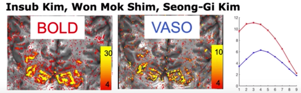
This figure is kindly provided by Insub Kim. - Reference data shown here
- Insub Kim, Won Mok Shim, and Seong Gi Kim are using layer-dependent VASO for orientation decoding across cortical depth.
- Max Planck Institute for Biological Cybernetics, Tuebingen, Germany:
- Jozien Goense used layer-dependent VASO in monkey visual cortex in areas of negative BOLD.

This figure is taken from Goense’s Neuron paper. - Data shown in Fig. 5 of this paper
- Jozien Goense used layer-dependent VASO in monkey visual cortex in areas of negative BOLD.
- University of Nottingham, Nottingham, UK:
- Rosa Panchuelo and Susan Francis are using ultra-high resolution VASO in order to map the sensory system.
- The grant is described here
- National Institute of Mental Health:
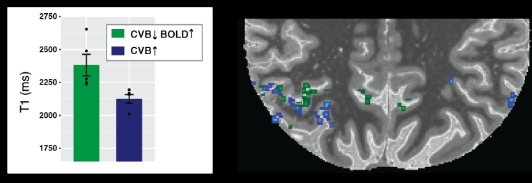
- Andrew Persichetti, Jason Avery, and Alex Martin are using layer-fMRI VASO to investigate the intra-cortical processing of imagined and executed motor actions.
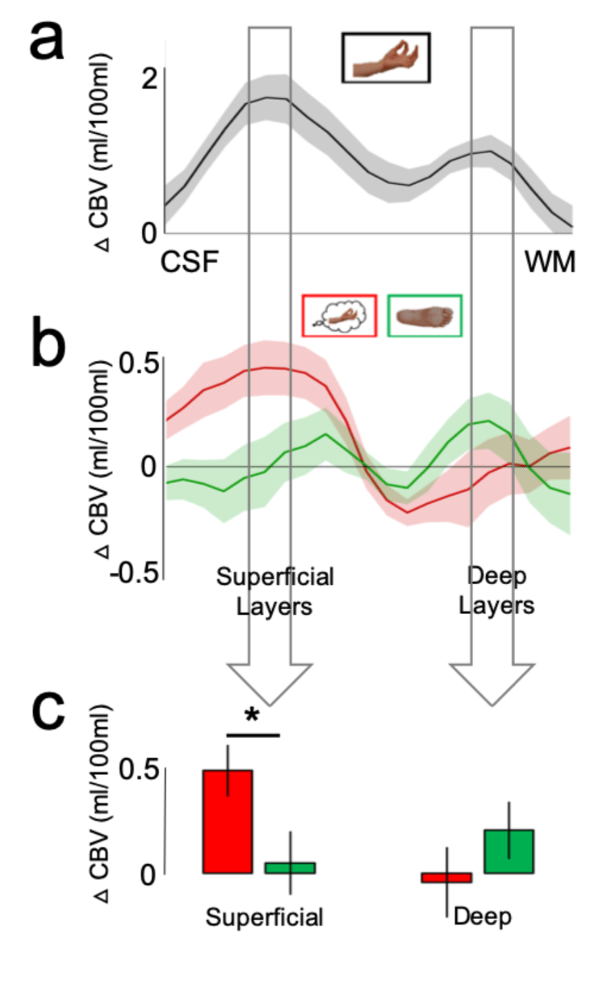
This figure is kindly provided by Andrew Persichetti. - Current Biology paper
- Andrew Persichetti, Jason Avery, and Alex Martin are using layer-fMRI VASO to investigate the intra-cortical processing of imagined and executed motor actions.
- CiNet, Osaka, Japan
- Ikuhiro Kida is using high-resolution VASO to investigate the neuro-vascular coupling features of fMRI.
- Sequence approved from SIEMENS in Feb 2019, ethical approval received in fall 2019.

Double stripe of tapping induced activity.
- NYU, New York USA
- University Magdeburg
- Esther Kuehn and Oliver Speck are piloting layer-fMRI VASO to investigate sensory-motor representations across cortical depth.

Figure credits: Esther Kuehn - Pilot study in June 2018
- Esther Kuehn and Oliver Speck are piloting layer-fMRI VASO to investigate sensory-motor representations across cortical depth.
- Christian Doppler Klinik, Salzburg
- Martin Kronbichler is investigating the usability of layer-dependent VASO at 3T.

Figure credits: Martin Kronbichler - Reference data shown here
- Martin Kronbichler is investigating the usability of layer-dependent VASO at 3T.
- NIPS, Okazaki, Japan
- Masaki Fukunaga is using layer-fMRI VASO in the sensory motor system, in the insual, and the visual cortex.
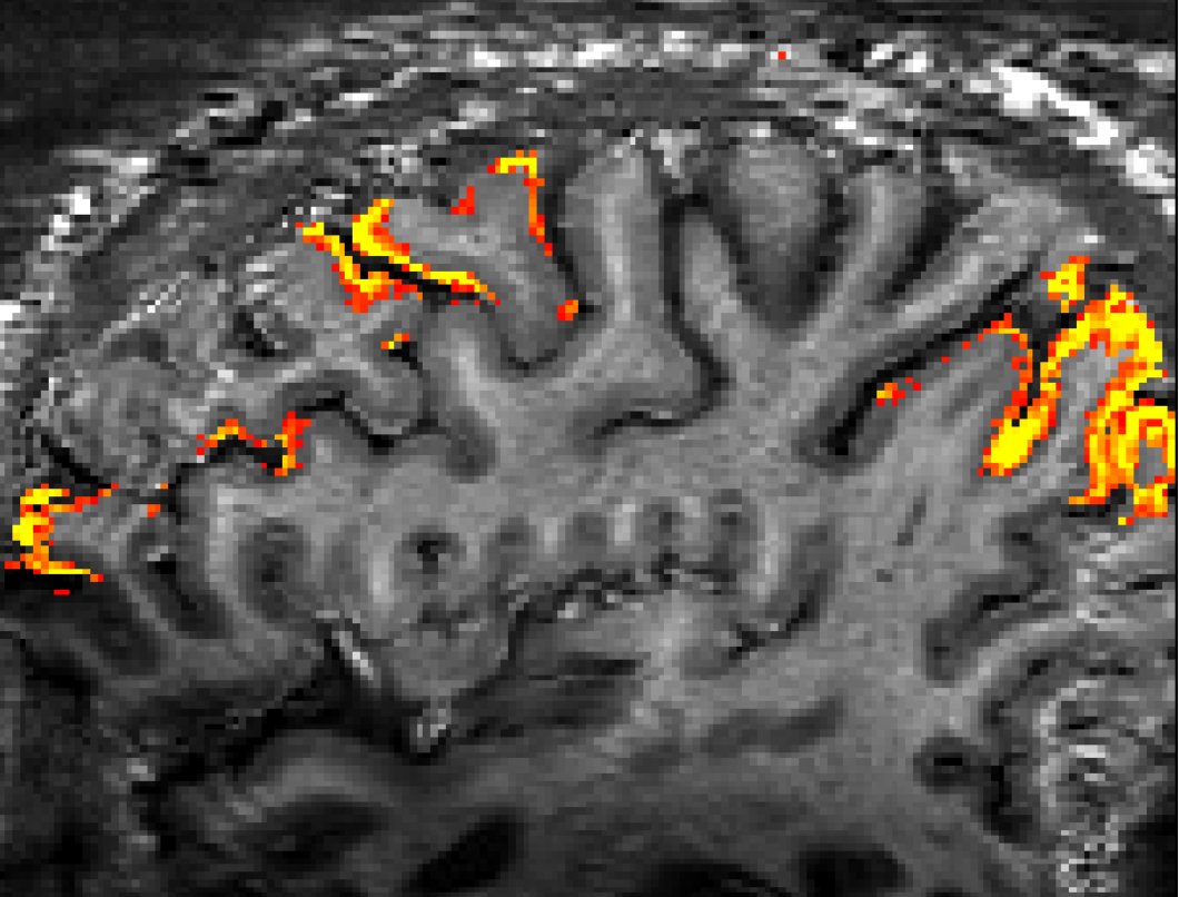
Figure credits: Masaki Fukunaga - Layer-fMRI VASO research agreement
- Masaki Fukunaga is using layer-fMRI VASO in the sensory motor system, in the insual, and the visual cortex.
- Okayama University Hospital, Japan
- Yinghua Yu is using layer-dependent VASO with predictive coding in the sensory system.
This figure is kindly provided by Yinghua Yu - Reference
- Yinghua Yu is using layer-dependent VASO with predictive coding in the sensory system.
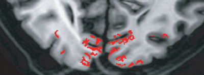
- Max-Delbrueck-Centrum, Berlin, Germany
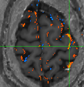
- Henning Reimann and Jurjen Heij are investigating layer-dependent processing of pain.
-

Example activation map from Henning Reimann at 0.8×0.8×0.7mm
- Zhejiang University, China
- Ruiliang Bai in the group of Anna Wang are using 7T VASO for high-resolution fMRI.
- Layer-fMRI research agreement is approved by SIEMENS
- Institute of Biophysics, Chinese Academy of Sciences, China
- Lab of Peng Zhang are using layer-fMRI VASO in humans at 7T
- Minnesota UHF workshop 2021
- University of Cambridge, UK
- Bingjiang Lyu and Chris Roger are working on the implementation of layer-fMRI VASO for application in speech fMRI.
- University of Cambridge, UK
- Catarina Rua and Zoe Kourtzi are setting up layer-dependent VASO in the visual cortex.
-

0.8mm VASO data from Cat Rua
- Oxford Centre for Functional MRI of the Brain, UK
- James Kolasinsky and Olivia Viessmann acquired high-resolution VASO with SMS readout for application in the somatosensory system.
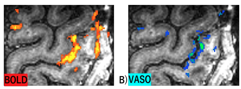
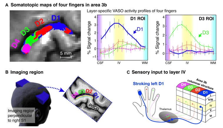
- Kennedy Krieger Institute, Johns Hopkins University, Baltimore, USA
- Jun Hua developed a high-resolution 7T VASO sequence and is applying it with working memory tasks in dementia patients.
This figure is taken from Hua’s MRM 2012 paper. - Reference
- Jun Hua developed a high-resolution 7T VASO sequence and is applying it with working memory tasks in dementia patients.
- Uniklinik Freiburg, Germany.
- Burak Akin and Ali Özen are acquiring layer-fMRI VASO at 3T with micro-stip RF-coils.
- Klinikum Erlangen, Germany
- Velentin Riedl, as collaborator from TUM, used VASO for quantitative fMRI at 7T.
- DZNE, Bonn, Germany:
- Ruediger Strinberg and Tony Stoecker implemented a VASO sequence with segmented 3D-EPI readout for SIEMENS VE systems.
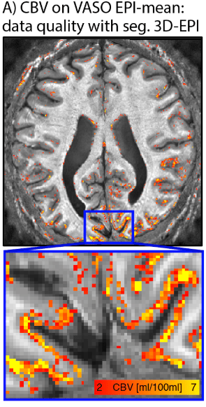
Data of this figure were acquired with Stirnberg’s sequence at the 7T Terra at NIH.
- Ruediger Strinberg and Tony Stoecker implemented a VASO sequence with segmented 3D-EPI readout for SIEMENS VE systems.
- Essen/Donders, Germany
- Victor Pfaffenrot and Oliver Kraft are using MAGEC VASO for layer-fMRI
- TUM, Munich, Germany
- Valentin Riedl is using VASO to avoid administration of contrast agents.
- Weizmann institute, Israel
- Edna Furman-Haran uses VE VASO on the Terra
- UIUC Illinois, USA
- Brad Sutton, Yuhui Chai aim to use VASO at the new Terra
- Berkeley, USA
- Prof. Feinberg is using layer-fMRI VASO on his Terra. This is to compare it with the results after the Terra is upgraded to the next-generation scanner.

- MPI Tuebingen, Germany,
- Vinod Kumar requested VASO for 9.4T scanning.
- Université Catholique de Louvain (UCL), Belgium
- Marco Barilari, and Remi Gau
- Neurococ, 2021, Liege
- Kings College London, UK
- Fraser Aitken
- Shanghai, Fudan Uni
- Deniz Vatansever for Terra and Prisma
- UT Dellas, USA, Southwestern
- Sina Aslan for Prisma
- Marseille, France
- Olivier Girard
- Bern, Switzerland
- Andrea Federspiel
- Showa, Tokyo, Japan
- Takashi Itahashi
- UC-Davis, USA
- Audrey Fan is using 3D-EPI VASO for vascular physiology mapping.
- London, Ontario, Canada
- Atena Akbari and Ravi Menon
- Rome, Italy
- Maria Guidi
- NTNU, Trondheim, Norway
- Desmond Tse and Pål Erik Goa are ramping up a layer-fMRI VASO grant for application in aging population.
- Reference
Forty papers showing data with layer-dependent VASO
- Donahue, M; Lu, H; Jones, C; Edden, R; Pekar, J; van Zijl, P. (2006). Theoretical and Experimental Investigation of the VASO Contrast Mechanism. Magnetic Resonance in Medicine.
- Jin, T; Kim, SG. (2008). Improved Cortical-Layer Specificity of Vascular Space Occupancy FMRI with Slab Inversion Relative to Spin-Echo BOLD at 9.4 T. NeuroImage.
- Goense, J; Merkle, H; Logothetis, N. (2012). High-Resolution FMRI Reveals Laminar Differences in Neurovascular Coupling between Positive and Negative BOLD Responses. Neuron.
- Bandettini, P. (2012). The BOLD Plot Thickens: Sign- and Layer-Dependent Hemodynamic Changes with Activation. Neuron.
- Huber, L; Ivanov, D; Krieger, S; Streicher, M; Mildner, T; Poser, B; Möller, H; Turner, R. (2014). Slab-Selective, BOLD-Corrected VASO at 7 Tesla Provides Measures of Cerebral Blood Volume Reactivity with High Signal-to-Noise Ratio. Magnetic Resonance in Medicine.
- Huber, L; Goense, J; Kennerley, A; Ivanov, D; Krieger, S; Lepsien, J; Trampel, R; Turner, R; Möller, H. (2014). Investigation of the Neurovascular Coupling in Positive and Negative BOLD Responses in Human Brain at 7T. NeuroImage.
- Huber, L; Goense, J; Kennerley, A; Trampel, R; Guidi, M; Reimer, E; Ivanov, D; Neef, N; Gauthier, C; Turner, R; Möller, H. (2015). Cortical Lamina-Dependent Blood Volume Changes in Human Brain at 7T. NeuroImage.
- Guidi, M; Huber, L; Lampe, L; Gauthier, C; Möller, H. (2016). Lamina-Dependent Calibrated BOLD Response in Human Primary Motor Cortex. NeuroImage.
- Huber, L; Ivanov, D; Guidi, M; Turner, R; Uludağ, K; Möller, H; Poser, B. (2016). Functional Cerebral Blood Volume Mapping with Simultaneous Multi-Slice Acquisition. NeuroImage.
- Donahue, M; Juttukonda, M; Watchmaker, J. (2017). Noise Concerns and Post-Processing Procedures in Cerebral Blood Flow (CBF) and Cerebral Blood Volume (CBV) Functional Magnetic Resonance Imaging. NeuroImage.
- Kazan, S; Huber, L; Flandin, G; Ivanov, D; Bandettini, P; Weiskopf, N. (2017). Physiological Basis of Vascular Autocalibration (VasA): Comparison to Hypercapnia Calibration Methods. Magnetic Resonance in Medicine.
- Huber, L; Handwerker, D;Jangraw, D; Chen, G; Hall, A; Stüber, C; Gonzalez-Castillo, J; Ivanov, D; Marrett, S; Guidi, M; Goense, J; Poser, B; Bandettini, P. (2017). High-Resolution CBV-FMRI Allows Mapping of Laminar Activity and Connectivity of Cortical Input and Output in Human M1. Neuron.
- Dumoulin, S. (2017). Layers of Neuroscience. Neuron.
- Poser, B; Setsompop, K. (2018). Pulse Sequences and Parallel Imaging for High Spatiotemporal Resolution MRI at Ultra-High Field. NeuroImage.
- Huber, L; Ivanov, D; Handwerker, D; Marrett, S; Guidi, M; Uludağ, K; Bandettini, P; Poser, B. (2018). Techniques for Blood Volume FMRI with VASO: From Low-Resolution Mapping towards Sub-Millimeter Layer-Dependent Applications. NeuroImage.
- Huber, L; Tse, D; Wiggins, C; Uludağ, K; Kashyap, S; Jangraw, D; Bandettini, P; Poser, B; Ivanov, D. (2018). Ultra-High Resolution Blood Volume FMRI and BOLD FMRI in Humans at 9.4T: Capabilities and Challenges. NeuroImage.
- Finn, E; Huber, L; Jangraw, D; Molfese, P; Bandettini, P. (2019). Layer-Dependent Activity in Human Prefrontal Cortex during Working Memory. Nature Neuroscience.
- Chai, Y; Li, L; Huber, L; Poser, B; Bandettini. (2019). Integrated VASO and Perfusion Contrast: A New Tool for Laminar Functional MRI. NeuroImage.
- Huber, L; Uludağ, K; Möller, H. (2019). Non-BOLD Contrast for Laminar FMRI in Humans: CBF, CBV, and CMRO2. NeuroImage.
- Persichetti, A; Avery, J; Huber, L; Merriam, E; Martin A. (2020). Layer-Specific Contributions to Imagined and Executed Hand Movements in Human Primary Motor Cortex. Current Biology.
- Yu, Y; Huber, L; Yang, J; Jangraw, D; Handwerker, A; Molfese, P; Chen, G; Ejima, Y; Wu, J; Bandettini, P. (2019). Layer-Specific Activation of Sensory Input and Predictive Feedback in the Human Primary Somatosensory Cortex. Science Advances.
- Yang, J; Yu, Y. (2019). “超高磁場・高精細レイヤー FMRI 技術による ヒト大脳皮質の層別活動の可視化.” Medical Science Digest 45(418): 418–21. http://hokuryukan-ns.co.jp/cms/books/medical-science-digest 2019年 6月臨時増刊号/.
- Huber, L; Finn, E; Handwerker, D; Bönstrup, M; Glen, D; Kashyap, S; Ivanov, D; Petridou, N; Marrett, S; Goense, J; Poser, B; Bandettini P. (2020). Sub-millimeter fMRI reveals multiple topographical digit representations that form action maps in human motor cortex. Neuroimage.
- Beckett, A; Dadakova, T; Townsend, J; Huber, L; Park, S; Feinberg, D. (2020). Comparison of BOLD and CBV using 3D EPI and 3D GRASE for cortical layer functional MRI at 7 T. Magnetic Resonance in Medicine.
- Guidi, M; Huber, L; Lampe, L; Merola, A; Ihle, K; & Möller, H. (2020). Cortical laminar resting‐state signal fluctuations scale with the hypercapnic blood oxygenation level‐dependent response. Human Brain Mapping.
- Huber, L; Finn, E; Chai, Y; Goebel, R; Stirnberg, R; Stöcker, T; Marrett, S; Uludag, Kim SG; Han, S; Bandettini, P; Poser B. (2021). Layer-dependent functional connectivity methods. Progress in Neurobiology.
- Huber, L; Poser, B; Kaas, A; Fear, E; Dresbach, S; Berwick, J; Goebel, R; Turner, R; Kennerley, A. (2021). Validating layer-specific VASO across species. NeuroImage.
- Oliveira, Í; Cai, Y; Hofstetter, S; Siero, J; van der Zwaag, W; Dumoulin, S. (2021). Comparing BOLD and VASO-CBV population receptive field estimates in human visual cortex. Neuroimage.
- Yu, Y; Huber, L; Yang, J; Fukunaga, M; Chai Y; Jangraw, D; Chen, G; Handwerker, D; Molfese, P; Ejima, Y; Sadato, N; Wu, J; Bandettini P. (2022). Layer-specific activation in human primary somatosensory cortex during tactile temporal prediction error processing. Neuroimage.
- Iamshchinina, P; Haenelt, D; Trampel, R; Weiskopf, N; Kaiser, D; Cichy, R. (2021). Benchmarking GE-BOLD, SE-BOLD, and SS-SI-VASO sequences for depth-dependent separation of feedforward and feedback signals in high-field MRI. bioRxiv.
- Akbari, A; Bollmann, S; Ali, T; & Barth, M. (2021). Modelling the depth-dependent VASO and BOLD responses in human primary visual cortex. bioRxiv.
- Yang, J; Huber, L; Yu, Y; Bandettini, P. (2021). Linking cortical circuit models to human cognition with laminar fMRI. Neuroscience & Biobehavioral Reviews.
- Huber, L; Kronbichler, L; Stirnberg, R; Ehses, P; Stöcker, T; Fernández-Cabello, S; Poser, B; Kronbichler, M. (2022). Evaluating the capabilities and challenges of layer-fMRI VASO at 3T. BioRxiv.
- Huber, L; Kassavetis, P; Gulban, O; Hallett, M; Horovitz, S. (2022). Laminar VASO fMRI in focal hand dystonia patients. BioRxiv.
- Koiso, K; Müller, A; Akamatsu, K; Dresbach, S; Wiggins, C; Gulban, O; Goebel, R; Miyawaki, Y; Poser, B; Huber L. (2022). Acquisition and processing methods of whole-brain layer-fMRI VASO and BOLD: The Kenshu dataset. BioRxiv.
- Liu, T. T., Fu, J. Z., Chai, Y., Japee, S., Chen, G., Ungerleider, L. G., & Merriam, E. P. (2022).
Layer-specific, retinotopically-diffuse modulation in human visual cortex in response to viewing emotionally expressive faces. Nature Communications, 13 (1), 6302. - Akbari, A., Gati, J. S., Zeman, P., Liem, B., & Menon, R. S. (2023). Layer Dependence of
Monocular and Binocular Responses in Human Ocular Dominance Columns at 7T using
VASO and BOLD. BioRxiv. - Faes, L. K., De Martino, F., & Huber, L. ( (2023). Cerebral blood volume sensitive layer-fMRI
in the human auditory cortex at 7T: Challenges and capabilities. PLOS ONE. - Dresbach, S., Huber, R., Gulban, O.F., Goebel, R. (2023). Fast layer-fMRI VASO with short stimuli and event-related designs at 7T. BioRxiv.
- Pizzuti, A., Huber, L., Gulban, O. F., Benitez-Andonegui, A., Peters, J., & Goebel, R. (2023). Imaging the columnar functional organization of human area MT+ to axis-of-motion stimuli using VASO at 7 Tesla. Cerebral Cortex.