Myths of basic EPI

Authors: Kenny (Chung) Kan1, Rรผdiger Strinberg2, and Renzo (Laurentius) Huber3

  1. Functional MRI Facility (FMRIF), NIMH, NIH, USA
  2. German Center for Neurodegenerative Diseases (DZNE), Germany
  3. Martino’s Center for Biomiedical imaging, Mass general Brigham, Harvard Medical School

What happens with the EPI sequence when I increase the TE? Which bandwidth should I use? What do I need to change to reduce PNS? There are many common misconceptions in basic EPI protocols. This blog post discusses the most common ones.

This post is a a continuation of highlighting basic acquisition features of EPI. See posts in GRAPPA, Regularization, Ghost correction, and 3rd order shim here.

Continue reading “Myths of basic EPI”

Sixth layer-fMRI dinner: layer-fMRI and the academia-industry relationship

On February 8th 2024, we will host the 8th virtual layer-fMRI dinner. As usual, it’s free, just follow the link below.

The 6th layer-fMRI dinner will focus the relationship of layer-fMRI academia and industry. Layer-fMRI basic research and industry have different goals, yet they help and facilitate one another. Without each other, each of our lives would be harder.

Continue reading “Sixth layer-fMRI dinner: layer-fMRI and the academia-industry relationship”

Second CMRR cdr workshop: Layer-fMRI analysis across pipelines

This post summarizes workshop of cortical depth resolved (cdr) fMRI, aka layer-fMRI Oct 10th-11th 2023. Official website is: https://sites.google.com/umn.edu/2023-minnesota-workshop/training/depth-resolved-layer-fmri. The slides, the data and the analysis skripts can be found here: https://doi.org/10.5281/zenodo.10004904.

The goal of this hands-on workshop was to:

  • Talk about the stuff that is not in the paper!
  • Show you the โ€œuglyโ€ parts of layer fMRI
  • Provide enough knowledge to allow analyzing layer data from beginning to end
  • Highlight the cool features of some of the most commonly used software

Best misheard sentence: “With Layer-fMRI, we have access to the cortical circus” (referring to circuit).

New established word inventions: “These data have been NORDICed

The workshop featured 4 independent pipelines of layer-fMRI analysis:

The data used here can be found on Zenodo: https://doi.org/10.5281/zenodo.10004904

Luca Vizioli: Introduction to the

Anna I Bล‚aลผejewska: FreeSurfer processing pipeline for laminar fMRI

Layer-fMRI analysis in LayNii and AFNI: Renzo Huber

Cheryl Olman: Nothing fancy: working with layer data with AFNI and FreeSurfer out-of-the-box

Fifth layer fMRI dinner: What can we learn about cortical layers using laminar fMRI?

On Dec 7th 2022, we will host the 5th virtual layer-fMRI dinner.

The board of the layer-fMRI dinner group has invited the following speakers to initiate discussions on the theme: Layer-fMRI signal origin: From neurons to vessels to BOLD.

  • Serge Dumoulin (NINS; Utrecht University): How population receptive field properties change across cortical laminae: from vision to cognition.
  • Anna Devor (Boston University): Layer-resolved imaging of resting CMRO2 in awake mice with phosphorescent O2 probes.
  • Lars Muckli (University of Glasgow): From man to non-human primates and mouse: Two top-down streams in different layers of retinotopic visual cortex (the ground truth).

Moderated by Luca Vizioli and Tyler Morgan 

The entire event will last for about 90 min (including discussion).

The meeting will be recorded and published on Youtube and embedded on this website by Dec 8, 2022. 

Everyone is welcome. No registration required. Zoom link:

https://layerfmri.page.link/Zoom

Los AngelesChicagoNew YorkUKEuropeBeijingSydney
6:008:009:0014:0015:0022:0023:00
Time Zones

Introduction

Serge Dumoulin
How population receptive field properties change across cortical laminae: from vision to cognition.

A key advantage brought by ultra-high field MRI at 7 Tesla and more is the possibility to increase the spatial resolution at which data is acquired, with little reduction in image quality. This opens a new set of opportunities for cognitive neuroscience, for example to probe how signals vary across cortical thickness and laminae. Here, I present recent work on computational modelling of population receptive field properties. I will discuss how they vary across cortical laminae, how these properties are influenced by attention and extend these protocols from primary visual cortex towards numerical cognition in association
cortex. I will also discuss some of the limitations and the potential of laminar imaging in human cortex.

Anna Devor
Layer-resolved imaging of resting CMRO2 in awake mice with phosphorescent O2 probes.

The cerebral cortex is organized in cortical layers that differ in their cellular density, composition, and wiring. Cortical laminar architecture is also readily revealed by staining for cytochrome oxidase โ€“ the last enzyme in the respiratory electron transport chain located in the inner mitochondrial membrane. It has been hypothesized that a high-density band of cytochrome oxidase in cortical layer IV reflects higher oxygen consumption under baseline (unstimulated) conditions. We tested the above hypothesis using direct measurements of the partial pressure of O2 (pO2) in cortical tissue by means of 2-photon phosphorescence lifetime microscopy (2PLM). We revisited our previously developed method for extraction of the cerebral metabolic rate of O2 (CMRO2) based on 2-photon pO2 measurements around diving arterioles and applied this method to estimate baseline CMRO2 in awake mice across cortical layers. Our results revealed a decrease in baseline CMRO2 from layer I to layer IV. This decrease of CMRO2 with cortical depth was paralleled by an increase in tissue oxygenation. Higher baseline oxygenation and cytochrome density in layer IV may serve as an O2 reserve during surges of neuronal activity or certain metabolically active brain
states rather than baseline energy needs.

Lars Muckli
From man to non-human primates and mouse: Two top-down streams in different layers of retinotopic visual cortex (the ground truth).

Using laminar fMRI, we identified two top-down processing streams. (1) One stream is for contextualizing visual input based on fast recurrent processing of contextual information and (2) a second top-down projection is used for visual imagery. Both top-down streams are based on non-direct geniculate input to visual cortex. To investigate the โ€˜ground-truthโ€™ of this top-down processing, collaborators in the Human Brain Project (HBP) conducted parallel experiments in monkeys and mice using microelectrode recording and two-photon calcium imaging. The contextual feedback effects in complex visual fields are fast, dependent on learning and are likely communicated by disinhibition in superficial layers of cortex.

Complex cognitive tasks are difficult to instruct in non-human primates and in rodents, but in humans we can see that top-down feedback processing is used for visual imagery, and also object comparison and navigation.

Discussion

Fourth layer fMRI dinner: Neurons โ†’ vessels โ†’ fMRI

On Oct 6th 2021, we aim to host the 4th virtual layer-fMRI dinner.

The board of the layer-fMRI dinner group has invited the following speakers to initiate discussions on the theme: Layer-fMRI signal origin: From neurons to vessels to BOLD.

  • Amir Shmuel (McGill): The complexity of lamina resolved neuronal activity, and the spatial specificity of BOLD, CBV, arterioles and venules responses: implications for planning and interpreting depth-dependent fMRI.
  • Jonathan Polimeni (MGH): Biophysical modeling for interpreting fMRI signals and relating them back to neuronal activity: contemplating the โ€œinverse problemโ€. 
  • Evelyn Lake (Yale): Leveraging simultaneous multi-modal fMRI and wide-field optical imaging to study functional brain networks.

Moderated by Luca Vizioli and Andrew Morgan 

Board:  Johanna Bergmann, Avery Berman, Saskia Bollmann, Denis Chaimow, Renzo Huber, Nils Nothnagel, Renรฉ Scheeringa, and Bianca van Kemenade.

The entire event will last for about 90 min (including discussion).

The meeting will be recorded and published on Youtube and embedded on this website by October 7th 2021. 

Everyone is welcome. No registration required. Zoom link: https://laminauts.page.link/meeting_channel 

Brisbane KoreaGermanyUK/UTCNew YorkMinnesotaSan Francisco
Oct 6thOct 6thOct 6thOct 6thOct 6thOct 6thOct 6th
11pm10pm3pm2pm9am8am6am

Introduction


Jonathan Polimeni (MGH)

Biophysical modeling for interpreting fMRI signals and relating them back to neuronal activity: contemplating the โ€œinverse problemโ€.

Abstract:

The ultimate limits of spatial and temporal resolution achievable by fMRI are dictated by neurovascular coupling, the mechanisms of blood flow regulation, and vascular architecture in the brain. While these limits are currently unknown, there is a rapidly growing body of evidence pointing to the ability of fMRI to distinguish site of activation across cerebral cortical depths, which can be used to infer the cortical layer or layers differentially engaged in specific tasks or functional networks. Because all fMRI signals currently in use are based on hemodynamics and hence are influenced by local vasculature, understanding how patterns of neural activity are transformed into the fMRI signals we measure can potentially aid not only in the interpretation of our data but also opens possibilities to better estimate the location (in space and time) and amplitude of the neural response from the fMRI responseโ€”to the extent that this transformation is โ€œinvertibleโ€.

Motivated by this, the goal of this presentation is to survey recent work towards building biophysical models of the fMRI signals to help with this interpretation, with a focus on models using realistic microvascular networks and dynamics based on optical imaging and microscopy data. These models are built on first principles and are described by meaningful anatomical and physiological parameters. I will present initial results demonstrating how these models can be used to predict well- known differences in the hemodynamic response across stimulus configurations and cortical depths. While these models are complex, and simulations are computationally intensive, they can be also used to help inform simpler โ€œlumpedโ€ models that are more practical for routine use, and are applicable to predicting various BOLD and non-BOLD fMRI contrasts.

Another goal of this presentation is to engage the laminar fMRI community and have an open discussion about the strengths and weaknesses of this modeling approach, consider these against other approaches to improve neural specificity in fMRI, and discuss how to combine this framework with advanced acquisitions and analysis methods towards our shared objective to measure neural activity across cortical layers with fMRI.

Video recording of Jonathan Polimeni’s presentation

Amir Shmuel, PhD | The Neuro - McGill University
Amir Shmuel (McGill)

The complexity of lamina resolved neuronal activity, and the spatial specificity of BOLD, CBV, arterioles and venules responses: implications for planning and interpreting depth-dependent fMRI

Video recording of Amir Shmuel’s presentation

Evelyn Lake (Yale)

Leveraging simultaneous multi-modal fMRI and wide-field optical imaging to study functional brain networks.

Video recording of Evelyn Lake’s presentation

Discussion

Video recording of the discussion session.

Layer-fMRI Analysis Project 2025

Conducted in October 13-14th 2025

Coordinator: Jonathan Polimeni, Renzo Huber, and Luca Vizioli

Training Faculty: Laurentius Huber, Rainer Goebel, Anna Izabella Blazejewska, Luca Vizioli, and Jonathan Polimeni

One dataset, many analyses: an overview of the diverse processing approaches in layer-fMRI.

The layer-dinner group would like to invite you to show us your analysis pipeline in a brief presentation at an upcoming โ€œLayer-fMRI dinnerโ€ in the Spring of 2022. The analysis of layer-fMRI data is challenging and not straightforwardly doable with standardized streamlines analysis packages. Most layer-fMRI groups have their own dedicated analysis solutions to account for layer-specific challenges. As such, the purpose of this event is:

  • To illustrate multiple layer analyses of members of the field, and for others to follow.
  • To highlight challenges of high-res and layer specific analysis.
  • To stimulate discussion about analysis challenges and solutions.
  • To give analysis developers a platform to advertise their analysis solutions.
  • To illustrate differences and similarities of pipelines.

Introduction by Jonathan Polimeni

Rainer Goebel: Brain Voyager

Lecture

Hands on

Renzo Huber: LayNii & AFNI

Lecture

Hands on:

Anna Blazejewska: Freesurfer

Lecture

Hand on instructions

For the BrainVoyager hands-on session by Rainer Goebel:

For the LayNii-Afnii hands-on session by Renzo Huber, please download and install the following programs:

For the FreeSurfer hands-on session by Anna Blazajewska, there are no prior preparations needed. You will get server access. You can access the server with a vnc viewer. 

  • Mac users can use the native VNC client.ย 
  • Windows users will need to install a client such as TightVNC Viewer (https://www.tightvnc.com).ย 
  • Instructions on how to use it are here.ย 

The data that we will work with are below:

Mirror as Gdrive (not recommended as it might run into download quota): https://drive.google.com/drive/folders/18joR_Kvil9OK13lZNMmbfOj_WYnETxqU?usp=sharing

Updates will follow

Third layer-fMRI dinner: Cognitive Models and Cortical Layers.

On April 20th 2021, the third virtual layer-fMRI took place. 120 (unique) attendees joined and discussed the connection between layer-fMRI and cognitive models.

This meeting is held as a succession of the first two virtual dinner in May 2020, and Sept 2020:

In this third event, it will be discussed how the layer-fMRI methodologies might be able to inform Cognitive models. The three speakers are researchers that are working to examine cognitive processes whose study is aided by understanding the structure and function of cortical layers. These cognitive processes could include memory, attention, learning, dreaming, language or cortical predictions (plus many, many more!)

Floris de Lange will give an overview of work done by his group to capture laminar fMRI activity changes in the visual cortex for prediction, attention and bottom-up input. Andrรฉ Bastos will present results of laminar LFP recordings and how feed-forward gamma-band and feedback alpha/beta band modulations help to understand cognitive effects including attention, working memory, and prediction processing. Michelle Moerel will talk about how computational models can be combined with laminar fMRI to understand human auditory processing. 

Below you find the important links of the virtual event. Embedded videos of the talks, discussions, and a summary of the hot topics are going to be added on the day after the event.

Continue reading “Third layer-fMRI dinner: Cognitive Models and Cortical Layers.”

Second Layer-fMRI dinner: Laminae in the brain; fMRI vs. electrophysiology

On Sept 28th 2020, the second virtual layer-fMRI event is scheduled.

This meeting is held as a succession of the first virtual dinner in May 2020: https://layerfmri.com/virtualevent1/

In this second event, it will be discussed how the research field can bridge the gap between layer-dependent activity measures that are obtained with fMRI and electrophysiology, respectively. Kamil Ugurbil will present the perspective of high resolution for human neuroscience, Lucia Melloni will present the perspective of depth-dependent electrophysiological recordings in humans, and Seong-Gi Kim will talk about the combination of both worlds, layer-fMRI and layer-dependent electrophysiological recordings.ย 

Below you find the important links of the the virtual event. Embedded videos of the talks, discussions, and a summary of the hot topics are going to be added on the day after the event.

Continue reading “Second Layer-fMRI dinner: Laminae in the brain; fMRI vs. electrophysiology”

First layer-fMRI Dinner: Layer-fMRI contrasts

On May 7th 2020, there was the first virtual layer-fMRI dinner event to discuss current issues in the field.

This meeting was held as a replacement of an originally planned layer-fMRI dinner at ISMRM and happened in succession of an earlier in-person layer-fMRI dinner in November 2019 (meeting minutes here).

Below you find the important links of the the virtual event, videos of the talks and discussions, and a summary of the hot topics that were discussed.

  • The meeting was organized by Luca Vizioli and Renzo Huber. And it was supported by CMRR (Essa Yacoub and Kamil Ugurbil) as well as the Maastricht-York partnership grant (PIs: Aneurin Kennerley and Renzo Huber).
  • There were 149 participants + 4 speaker!
  • The layerfMRI slack-channel of the network has been opened to everyone and can be joined here: https://tinyurl.com/cdrfmri1.
  • The content of the next meeting will be determined by results of the survey here: https://layerfmri.page.link/meeting_survey. The meeting is scheduled for early July as of May 8th, it looks like most people prefer to talk about analysis challenges.

Hot topics that were discussed

  • How do we estimate sensitivity and specificity of a sequence?
  • Validations that layer-specific fMRI signals are explainable by electrophysiology are necessary.
  • How important is it to consider arterial artifacts for layer-fMRI signal interpretation?
  • How are inflow effects considered in different layer-fMRI readout schemes?
  • Can maps of physiological noise be helpful for segmentation and/or registration?
  • What do maps of non-gaussian noise represent?
  • The major limitation of layer-fMRI is still the resolution! Can we go to smaller voxels? How?
  • How can we make use to layer-fMRI in non-primary areas? And how applicable is it?
  • How can we make use of layer-fMRI in pathology? E.g. vascular diseases.
  • The combination of experimental setup and acquisition contrast is important.
  • Layer-fMRI is depth-dependent fMRI.

Talks and Discussion of the virtual event

All slides can be downloaded here: https://doi.org/10.5281/zenodo.3874364

Next meeting


The next virtual layer-fMRI dinner will tentatively be on September 28th (Europe and America) and September 29th (in Asia) on the topic on layer-fMRI vs. electrophysiology. with speakers including Kamil Ugurbil and Seong-Gi Kim.

2019 Minnesota workshop on Cortical Depth-Resolved fMRI Methods

This post summarizes the presentation, tutorials, and discussions of the 2019 UHF Minnesota Workshop on Cortical Depth-Resolved fMRI Methods, Nov 12th-Nov 13th.

OrganiserCheryl Olman
PresentersAlessio FracassoNatalia PetridouJonathan PolimeniKamil UludagTim van Mourik, and Renzo Huber

Terminology consensus ๐Ÿ™‚

  • Layerification: The process of assigning depth-values to each voxel.
  • Lettuce Head: Levelset with acoustic noise.
  • Partial Volume: Ill-defined term for partial coverage. A field of view that is smaller than what freesurfer considers as โ€œwhole brainโ€.

Workshop Content

Cheryl Olman: Introductions and general outline: 

Jonathan Polimeni: Overview of laminar fMRI best practices and current challenges

Part 1:

Part 2:

Hot Topic discussions included: 

  • How many cytoarchitectonically-defined layers are there, is 6 really a good number?
  • How should we estimate the PSF?
  • Resolution losses from resampling should be kept as small as possible. Viable strategies include: 1.) working in upsampled space, 2.) combining (concatenating) all transformations into one single transformation, 3.) using adequate interpolation functions. 
  • Layer smoothing can be helpful to depict layer activation features, but they should always be accompanied with unsmoothed maps. Otherwise it can result in circularity. 
  • โ€œshiftingโ€ the cortical depth based on the functional baseline signal a la Peter Koopmanns might introduce circularity. Tim: this approach is debunked. 
  • The accuracy of the surfaces (segmentation lines) is significantly higher than the voxel resolution. This is possible with predefined assumptions of signal intensities of GM and WM and a partial voluming model. -> higher resolution of the anatomy helps to improve the accuracy. However, itโ€™s more important to keep the SNR up.

Renzo Huber: Hands on scanning at 7T scanning: Optimizing an EPI acquisition

Tim van Mourik, Using GIRAFFE to set up analysis pipeline (boundary-based registration)

Part 1:

Part 2:

Alessio Fracasso: Hands on analysis: Segmentation and layerification without surfaces

  • Digital capture failed; weโ€™re working on creating a replacement
  • Hot topic discussion: 
    • The presented pipeline estimates layers with level-sets, without surfaces in voxel space.

fMRI contrasts:  GE-BOLD, SE-BOLD and non-BOLD

  • Natalia Petridou: GE, SE, GRASE
    • Hot Topic discussion:
      • GRASE has the advantages of both GE-BOLD and SE-BOLD, or does GRASE have the disadvantages of both GE-BOLD and SE-BOLD?
      • Different layers and different contrasts have different timing response functions.
  • Renzo Huber: non-BOLD (VASO, ASL โ€ฆ)
    • Hot topic discussions: 
      • There is no clear winner of sequences. Sequence comparisons are never fair. 

Kamil Uludag: T1-weighted EPI and laminar BOLD response modeling

  • Hot topic discussions: 
    • There is no easy ground-truth of tissue type segmentation. When comparing methods, one needs to look at both approaches.
    • -> taking the difference between task conditions and using the layer-dependent activation difference for neuroscience interpretations is not adequate <- This does not mean that previous studies, who did this are necessarily wrong.
    • The vein size difference across layers can be incorporated in the model as CBV. 
    • The vascular deconvolution method might come along with noise-amplification. When you have unreliable data quality to begin with, the deconvolution model might make more problems than it solves.
    • The surprising CBF profiles are in agreement with previous studies from Ingo Marquardt and from electrophysiology.

Natalia Petridou: 3D-EPI 

  • Hot topic discussions: 
    • It is not so straightforward to correct for physiological noise when you have long readouts in 3D-EPI. The most appropriate approach is to take it as a snapshot acquisition at k-space center. 
    • K-space based approaches like RetroKCor might be more appropriate for 3D-EPI 
    • It is not clear, why the physiological noise should become less severe in the thermal noise dominated regime? Itโ€™s more important how big the physiological noise is with respect to the BOLD magnitude? Itโ€™s less important how big the physiological noise it with respect to the thermal noise?
    • Offline-discussion with Natalia Petridou: Motion is the single biggest limitation in high-res fMRI.  The most effective way to minimize motion is to engage the participant. E.g. reward, if motion is low. E.g. penalty-based longer time in the scanner (repetition of runs), when motion is large.

Renzo Huber: Hands on Analysis: Layerification with LAYNII

  • Recording Part 1:
  • Recording Part 2:
  • Hot topic discussions:
    • How many layers should be extracted?
      • Renzo Huber: extract as many layers as possible (potentially after upsampling).
      • Tim Van Mourik: extract as many layers as independent samples across cortical depth.
      • Consensus among all: the least subjective choice is to have as many layers as voxels. These layers are sparse and non-independent.
      • Consensus among all: any number of layers is ok. 
    • Which interpolation function should one use to work in upsampled space
      • Consensus is that nearest neighbor is not adequate because it assumes that the signal would be equally distributed within the voxel.
      • Most adequate interpolation function would be zero-filling in k-space, which corresponds to sinc-interpolation in image space.
      • Consensus among all: if the result depends on the interpolation function, we shouldnโ€™t trust the result to begin with.
    • How should we do statistics with sparse and non-independent voxel sampling across depth?
      • Consensus among all: This is an unsolved problem. We donโ€™t even know which signal magnitude to trust. Thus, itโ€™s even less clear, how to do statistics with it. 
    • Why is it such an obstacle, if a software package has dependencies to GSL. Future versions of LAYNII should not be dependent on it?
    • It shouldnโ€™t be so hard to make LAYNII compatible with nii.gz, Future versions should be able to read nii.gz. 
    • The advantages and disadvantages of equi-volume and equi-distance approaches where discussed. Renzo advises to use equi-distance. While it contains negligible biases with respect to the cyto-layers, it does not come along with noise amplification as equi-volume.

Cheryl Olman:  A โ€œcompleteโ€ scanning session (MP2RAGE, some 3D GE EPI comparisons, T1-EPI)

  • Hot topic discussions: 
    • Setup of T1-EPI, how to analyze it correctly?

Alessio Fracasso: Surface-based visualizations/partial brain segmentations

  • Hot topic discussions: 
    • Looking at EPI data in anatomical space. 
    • How to minimize curvature bias of segmentation -> higher resolution. 

Cheryl Olman: Discussion sessions throughout the workshop: 

  • What are the most important challenges of layer-fMRI? 
    • Nominal resolution is not the same as effective resolution
    • There is no ground truth of quantifying the effective resolution (acquisition, biological, resampling).
    • Layerification is hard with distortion and registration challenges. 
    • Anatomical segmentation
    • The biggest challenges were obtained in survey from ISMRM study group: https://doi.org/10.7490/f1000research.1115658.1
  • What should every manuscript include?
    • All standard sequence parameters must be reported. Furthermore, parameters of echo-train length, partial Fourier etc. should be mentioned too.
    • Images of EPI data quality, e.g. representative tSNR maps, activity maps in native EPI space. 
    • Data of segmentation quality and registration quality should be shared.

Cheryl Olman: Wrap-up discussions:

  • Where do we want to host workshop content?
  • How to continue discussions: 
    • Active members of the community (who know how to use SLACK) will continue discussions on the SLACK workspace depthresolvedfmri.slack.com, This channel will be open to every layer-enthusiast (in an invitation basis). If you have not received an invite yet, please contact us. 
    • Parts of the discussions will be mirrored on layerfMRI.com, including:
      • Meeting minutes
      • Continuously updated list of layer-fMRI papers (with a focus on human fMRI).
      • List of job opportunities in layer-fMRI.
      • List of layer-fMRI abstracts of current conferences. 
  • Do we want a white paper on a set of QC metrics (tSNR in ROI, true image resolution in RO/PE/SL directions, ?) that can be used to compare acquisitions?
    • Response from all: Maybe 
    • Cheryl will contact the field about this soon.
    • As opposed to the field of ASL, we donโ€™t have a 20 year ongoing discussion or well-established agreed-upon standards. Thus, it might be challenging.  But there will probably be a basic set of agreed best practices.
  • Future satellite meetings: We want to keep organizing satellite meetings and informal meet-ups at conferences like ISMRM (Who volunteers? Who will attend? (e.g. Renzo and Luca?)), OHBM (-> Amir Shmuel), SfN and the BRAIN Investigators meeting (-> Sean Marrett).

layer-fMRI seminar

DateSpeakertopic
April 13th 2026
March 30th 2026Suvi Hรคkkinen
Functional imaging of hippocampal layers using
VASO on the Next Generation (NexGen) 7T
March 16th
2026
Wietske van der ZwaagMesoscale (f)MRI of the Cerebellum
March 2nd
2026
Alard RoebroeckImaging layer specific meso- and microstructure in the human brain with ex vivo MRI and light sheet microscopy
Feb 16th
2026
Holiday (Presidents Day)
Feb 2nd
2026
Dana RamadanMacrovascular contributions to resting-state fMRI signals: A comparison between EPI and bSSFP at 9.4 Tesla
Jan 19th
2026
Tony CarricarteLayer-specific spatiotemporal dynamics of feedforward and feedback in human visual object perception
Christmas break
Dec 8th
2025
Saskia BollmannTowards Quantitative Analysis of Vascular Characteristics in the Human Brain
Nov 24th
2025
Alejandro Monreallayer-fMRI spirals: VASO fMRI spirals, 11.7T fMRI spirals, etc.
Nov 10th
2025
Omer Faruk GulbanMeso-vessel imaging with 7 T MRI: Linking anatomy and function in living humans
Oct 27th
2025
Renzo Huber: PNS Optimized Pulses for EPI (POPE): Simple adjustment to gradient pulse shape for practical high-resolution fMRI. 
https://layerfmri.page.link/POPE
Divya Varadarajan: SurfScribe: Cortical surfaceโ€“driven automated online slice prescription applied to ultra-high-resolution vascular MRI
Renzo Huber: LN2_FRISGO: A software solution for artifact mitigation in fast high-resolution fMRI. https://layerfmri.page.link/LN2_FRISGO
Alessandra Pizzuti: Layer-fmri at 0.4 mm isotropic meets vascular mapping (0.35 mm iso): Partners or Confounders?
Oct 13th
2025
CMRR workshopHands on layer-fMRI analysis course with LayNii, FreeSurfer, and BrainVoyager. By Luca Vizioli, Renzo Huber, Jonathan Polimeni, Rainer Goebel.
Sept 29th
2025
Shahin NasrMesoscale cortical plasticity in humans revealed by high-resolution functional MRI
Sept 15th
2025
Amelia StromQuantification of cerebral cortical displacement driven by visual stimulation using motion-encoded stimulated-echo EPI at 7T
Sept 1st
2025
Labor DayNO MEETING
Aug 18th
2025
Sharif KronemerThe human brain mechanisms of afterimages: From networks to cortical layers
Aug 4th
2025
Marianna SchmidtMesoscale functional connectivity within the human primary visual cortex
June/Julysummer breakNO MEETING
May 27thFaruk Gulbanโ Faster, Finer, Higher, Larger with LayNii IDA: Meso Veins Meet Layer fMRI in High-Speed Data Exploration for the New Mesoscopic Era
May 12thcanceledISMRM Hawaii
April 28thStephanie, Khazar, Renzo, MarcoISMRM mock presentations: dynamic CSF volume imaging, 3rd order shim, layer-fMRI in hippocampus, layer-toolbox
April 14thElisa ZamboniMapping Curvature Domains in Human V4 Using CBV-Sensitive Layer-fMRI at 3T
March 31stcanceledISMRM workshop Annapolis
March 17thGabi LohmannCylarim: a new tool for laminar-specific fMRI analysis
March 3rdFraser Aitkenlayer-fMRI and epilepsy
Feb 17th canceledHoliday (Washingtons birthday)
Feb 3rdRenzo HuberUpdates on EPI artifact mitigations: towards whole brain layer-fMRI with faster TRs
Jan 22nd
10:30am
Emma BrouwerCerebellar imaging using 7T MRI
Winter break
Nov 27thLonike Faeslayer-fMRI in Auditory cortex: VASO sequence, NORDIC denoising, odd-ball tasks
Nov 13thKhazar AhmadiDeep dive into hippocampus from a laminar perspective
Oct 30thISMRM abstracts
Oct 16thGrace Edwardslayer-fMRI language effects in V1
Oct 2nd Yuhui ChaiBlood-nulling versus tissue-suppression: Enhancing integrated VASO and perfusion (VAPER) contrast for laminar fMRI
Sept 18thTyler MorganInvestigating neural responses using fast, non-selective MRI
Sept 4thDaniel HaeneltUnderstanding biases in functional magnetic resonance imaging
Aug 21st Praveen ValsalaAccelerating bSSFP-fMRI with Spiral Readouts
Aug 7thRenzo, Joelle, Tyler7TANA highlights
July 31stRenzo Huber7T initiatives across sites
July 25th
Thursday
Yulia Lazarova
July 22th
Monday
Alessandra Pizzuti
July 10thOHBM debriefingRenzo on holidays
Jun 26thOHBMcanceled
June 12th Faruk GulbanPhase Jolt fMRI
May 29th Sebastian Dresbach Laminar CBV and BOLD response-characteristics over space and time in human primary somatosensory cortex at 7T
May 15th ISMRM post discusion
May 1st, 2024
B1D55
ISMRM mock presentationsBurak (Layer-ReHo), Renzo (lower brain structures)
April 31st 2024
virtual
ISMRM mock presentationsTyler: DIANA & VAPER
April 17th,
2024
B1D55
ISMRM preparations Kenny (T1234), Lasse (NORDIC)
April 3rd, 2024
B1D55
 ISMRM mock presentationsRenzo (Motion symposium)
March 20th, 2024 B1D55Alessandra Pizzuti, about motion quartet confirmed
March 6th, 2024
B1D55
Marco Barilari: Characterizing multisensory integration and cross-modal plasticity in the cortex layers using VASO at high-res (7T)hybrid
Feb 21st,2024,Yuhui Chai Improving laminar fMRI specificity by reducing macrovascular bias revealed by respiration effects
Feb 7th, 2024 
virtual
Dongho Kim on Attention effects in human S1DIFFERENT TIME: 9:30am, virtual only
Jan 24th, 2024, 
virtual
SE-BOLD GE-BOLD, Face perception in V1 and ventralLuca Vizioli, virtual only
Jan 10th, 2024Yuhui Chai: Improving laminar fMRI specificity by reducing macrovascular bias revealed by respiration effects
Dec 27th canceledChristmas break
Dec 13thBurak Akinlayer-fMRI patch analysis to look for instantaneous layer profiles
Nov 29th Ethan Buch:layer-fMRI VASO on motor learning (Leo Cohenโ€™s lab)layer-fMRI VASO on motor learning (Leo Cohenโ€™s lab)
Nov 15th Canceled due to SFN in DC
Nov 1stISMRM abstracts 10:30-11:30
Oct 18thKenny Chung: T1234 EPI.Discussions of what we will submit on Nov 8th.10:30-11:30 
Oct 4th CanceledCanceled due to Boston Workshop : https://education.martinos.org/workshop-on-laminar-fmri/ 
Sept 20thRehearsal talks for layer-FMRI talks in Boston10:30-11:30
Sept 6thGrant HartungVAN layersTitle: Capillary density induces โ€œmicrovascular biasesโ€ in layer-fMRI BOLD: insights from realistic vascular modeling
Aug 23rdCanceled due to holidays
Aug 9th OHBM debriefing?
July 26th Canceled due to OHBM
July 12th Jiajia presents Layer-specific finger representations in human area 3b, abstract
June 28th high -resolution VASO in focal hand dystonia patientsSilvina HorovitzNote that this meeting will be 30 min earlier.
June 14th ISMRM post discussionRenzo Huber
May 31stISMRM rehearsal presentationsRenzo will present 9 min talk on fuzzy ripples and 20 min educational lecture on recent advances in the field of layer-fmri. Yuhui presented the VAPER connectivity.
May 17thDaniel ZaldivarLayer dependent changes of neural activity underlying laminar fMRI 
May 3rdFarukTutorial on ITK snap segmentation, QnATo video  https://youtu.be/tIuKG3rtVk4
April 19th TylerCortical-subcortical connection overview
April 5th Sam Audrain and Andrew Persicetti Talking about their endeavors to capture layer-fMRI in some of the most inferior parts of the cortex.
March 22ndEli Meriam Informal discussion of future layer fMRI study on texture processing in V1/V2
March 8th DIANA with Aneurin KennerleyTyler and Renzo are considering inviting Aneurin Kennerley to present his results with human line scanning at 3T. Aneurin confirmed
Feb 22ndAtena Akbariphase-regression and VASO for layer-fMRI in ocular dominance columns
Feb 8th Jun Hua Jun Hua will present his work on layer-fMRI with memory encoding in the entorhinal cortex.
Jan 25th 2023Sohuyn Hanโ€™s papers on Spin echo Han S, Eun S, Cho H, Uludaวง K, Kim SG. Improved laminar specificity and sensitivity by combining SE and GE BOLD signals. NeuroImage. 2022 www.doi.org/10.1016/j.neuroimage.2022.119675  Mini talk summary: https://youtu.be/ebDwcmcP4hw 
Han SH, Eun S, Cho HJ, Uludaวง K, Kim SG. Improvement of sensitivity and specificity for laminar BOLD fMRI with double spin-echo EPI in humans at 7 T. NeuroImage. 2021 https://doi.org/10.1016/j.neuroimage.2021.118435
Jan 11th 2023CanceledSohuyn Hanโ€™s papers on Spin echo Canceled Han S, Eun S, Cho H, Uludaวง K, Kim SG. Improved laminar specificity and sensitivity by combining SE and GE BOLD signals. NeuroImage. 2022 www.doi.org/10.1016/j.neuroimage.2022.119675  Mini talk summary: https://youtu.be/ebDwcmcP4hw 
Han SH, Eun S, Cho HJ, Uludaวง K, Kim SG. Improvement of sensitivity and specificity for laminar BOLD fMRI with double spin-echo EPI in humans at 7 T. NeuroImage. 2021 https://doi.org/10.1016/j.neuroimage.2021.118435
Dec 28th 2022Probably canceled with people on holidays?
Dec 14th 2022One week before the OHBM deadline.Discussion of abstracts to be submitted 
Nov 30th 2022 Yuhui presents connectivity results Layer-specific functional connectivity with 3D VAPER fMRI http://submissions.mirasmart.com/ISMRM2023/ViewSubmissionPublic.aspx?sei=4oW3ybRR7
Nov 2nd 2022canceled
October 19th 2022Erwin Hahn Lecturehttps://youtube.com/playlist?list=PLuA0pYRPZ4uAvC2uIHggzyQHveRrHLosx

This page gives an overview of the bi-weekly meetings of layer-fMRI researchers at NIH/Maastricht/MGH and and friends. We meet every other week on Monday 10:00am (EST).

Email reminders are sent via the listserv: layer-fmri@researchlist.partners.org. You can subscribe by sending an email to subscribe-layer_fmri@researchlist.partners.org or contact me, and I can add you to the list.

We use this conference channel: https://mgb-org.zoom.us/my/layerfmri.

Some presentations are recorded. The Youtube channel of all recordings is here and embedded below (select video out of playlist with he button on the top right).

Playlist of all presentations:

Agenda

Continue reading “layer-fMRI seminar”

Meeting minutes of the virtual layer-fMRI event #1

On May 7th 2020, there was a virtual layerfMRI event to discuss current issues in the field.

This meeting was held as a replacement of an originally planned layer-fMRI dinner at ISMRM and happened in succession of an earlier in-person layerfMRI workshop in November 2019 (meeting minutes here).

Below you find the important links of the the virtual event, videos of the talks and discussions, and a summary of the hot topics that were discussed.

Continue reading “Meeting minutes of the virtual layer-fMRI event #1”

MP2RAGE: challenges and artifacts of its use for layer-fMRI

The MP2RAGE sequence is very popular for 7T anatomical imaging and is very commonly used to acquire 0.7-1 mm resolution whole brain anatomical reference data. Aside of this common application, it can also be very helpful for layer-fMRI studies to obtain even higher resolution T1 maps in the range of 0.5mm iso. However, when optimizing MP2RAGE sequence parameters for layer-fMRI studies, there are a few things that might be helpful to keep in mind.

In this post, I would like to discuss the challenges of using the popular MP2RAGE sequence in layer-fMRI studies. Specifically I will discuss challenges/features regarding:

Continue reading “MP2RAGE: challenges and artifacts of its use for layer-fMRI”

Quick example of cortical unfolding in LAYNII

Update June 2021: There is a new and more comprehensive 3D-supported LayNii Program for cortical unfolding. Please see https://thingsonthings.org/ln2_multilaterate/

The blog post below is kept for archiving purposes:ย 

In this Blog post, I seek to describe a quick example of how to analyse high-resolution data across layers and columns with LAYNII.

Continue reading “Quick example of cortical unfolding in LAYNII”

Negative Voxels in VASO

In the last years, I and multiple other VASO users have encountered many occasions of voxels that show negative CBV change (positive VASO signal change) while the BOLD suggests that the activation should be positive. In this blog post, I want to list potential sources of this surprising effect.

Continue reading “Negative Voxels in VASO”

3D-printing nii data

In this blog post, I want to write about pipelines on how to prepare Nifti-brain data and make them printable by a 3D-printer.

Two pipelines are shown. One pipeline describes the 3D-printing the cortical folding structure that is estimated with Freesurfer and subsequently corrected with Meshlab. And another pipeline describes how you can 3D-print any binary nii-volume by using the AFNI-program IsoSurface and correct the output with netfabb.ย  Continue reading “3D-printing nii data”

2018 layer-fMRI meeting as part of the ISMRM study group ‘current issues in brain function’

Meeting minutes of Study group on brain functions business meeting:

15:30-16:30 in W07, June 21st, 2018, ISMRM, Paris:

Chunlei Liu presents the talk: โ€œUltra-high resolution fMRI – from hardware to pulse sequences to the human brain – and vice-versa. A 7T BRAIN Initiative Projectโ€ per pro David Feinberg.

In the subsequent Q/A period it is discussed that the current gradient design will be โ€œasymmetricโ€ and that the target parallel-imaging acceleration factor will be 20-30.

Past Chair Essa Yacoub presents the study group statistics:

There are many more trainees than full members.

Natalia Petridou presents the Trainee abstract awards:

  • #1 Adam Yamamoto for the abstract entitled: Intra-operative acquisition of sensorimotor fMRI during glioma resection: evaluation of feasibility and clinical applicability.
  • #2 Domenic Cerri for the abstract entitles: Mechanisms underlying negative fMRI response in the striatum. Since Dominic could not make it, he sent one of his collogues to receive the price for him.
  • #3 Yi-Tien Li for the abstract entitled: Inter-Regional BOLD Latency after Vascular Reactivity Calibration is Correlated to Reaction Time.

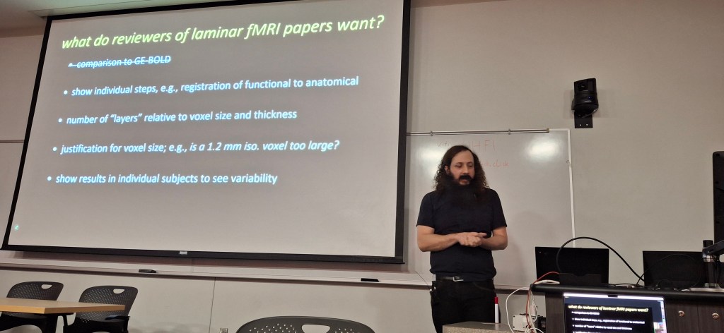
Renzo Huber presents the results of the survey among the study group members โ€œBiggest challenges of high-resolution fMRIโ€. The slides can be downloaded here: https://doi.org/10.7490/f1000research.1115658.1

The attendees of the business meeting commented on the biggest challenges of high-resolution fMRI and the issues that could be discussed in a future virtual study group meeting.

ยท   Unknown (to me) senior study group member: At very high resolutions, the elastography of the brain might become a limit of submillimeter fMRI. This comment might be related to the Tweet from NIH Director Francis Collins that went viral on social media earlier that day: https://twitter.com/NIHDirector/status/1009793641550893056 (with amplified motion).

ยท   Ravi Menon: Motion is a big limitation of sub-millimeter fMRI.

ยท   Cheryl Olman: Every person/group uses a different analysis pipeline. It would be helpful to have a synthetic standard dataset that is open to everyone. Every person could play around with it and test the quality of various evaluation pipelines.

ยท   Hanzhang Lu: The virtual study group meeting would lose the interest of many study group members if the focus is only of 7T imaging. It would be helpful to also discuss the spatial specificity limitations at 3T too.

ยท   Shella Keilholz: Since there are so many trainee members, they should mention what they need.

ยท   Olivia Stanley (โ€œencouragedโ€ from Ravi Menon):

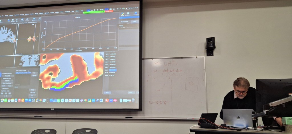
  1. Trainees want to know what segmentation tools are out there and how to use them.
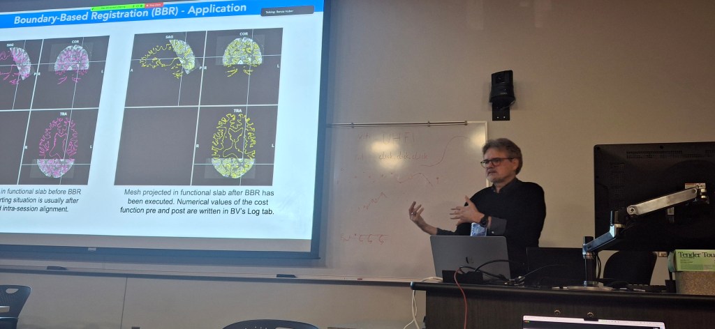
  2. Trainees want more information on registration methods.
  3. Trainees would benefit from a tutorial on how to share code and how to use shared code.

ยท   Ravi Menon: So many people are sharing their code on various platforms. It would be helpful to have an online repository of repositories.

ยท   Essa Yacoub: It will be hard to standardize analysis pipelines because the data and the acquisition hardware is not standardized.

ยท   Trainee member (not known to me): The analysis of sub-millimeter fMRI takes a lot of computational recourses. Trainees can be limited by this.

ยท   Nikolaus Weiskopf: The image reconstruction can be a black box. It is not straight-forward to share pipelines of the Image reconstruction.

ยท   Nikolaus Weiskopf: Data-sharing becomes more difficult in the EU because of increasingly stricter privacy protection regulations.

ยท   Natalia Petridou: Data sharing is increasingly encouraged from the funding agencies and the journals.

ยท   James Pekar: The field of sub-millimeter fMRI stands on the shoulder of decades of preceding work from volunteers and people with passion. This study group can help todays volunteers and people with passion to increase their visibility.

2015: The Nijmegen Layers Analysis Meeting

On October 22nd and 23rd 2015, David Norris hosted the first layer-fMRI workshop in Nijmegen. This workshop was focusing on hands-on analysis questions in layer-fMRI. Attendees were asked to share example data across vendors and acquisition methods. The agenda contained open discussions on the following questions:

  1. What data quality can we expect?
  2. Image registration
  3. Defining and determining cortical surfaces
  4. The equi-volume principle (Bok)
  5. How to determine layer surfaces and how many layers should I model
  6. Will it ever be possible to really know the depths of the histological layers in vivo?
  7. What are the realistic benefits of high resolution and layer specific fMRI?

Summary of the Meeting discussions:

Session on Image Registration

Coordinators: Sue Francis, Rosa Panchuelo (Nottingham)

  • Maastricht group mentioned that it is beneficial for layer0fMRI to use small FOVs. Best is to use IR-EPI for segmentation. Mean EPI can also be used, but the contrast between GM/WM is not as good. Acquire a scan with inverted PE direction. Preferably register anatomy to EPI, not the other way around: we want to leave the functional scans as untouched as possible.
  • It is mentioned that RETROICOR would beneficial at resolution of 1mm isotropic for 3D EPI.
  • It is furthermore suggested to use GLM noise (noise mask + PCA of noise).
  • Rosa Panchuelo mentions that AFNI is the best tool for motion correction.
  • Tim van Mourik recommends affine registration with boundary-based cost functions (FSL or FreeSurfer).
  • Kamil Uludag: we need a more objective way of assessing the quality of registration, people do it by eye at the moment.

Session on Cortical Surfaces

Coordinator: Pierre-Louis (Pilou) Bazin and Sriranga (Sri) Kashyap

  • Pilou showed how to use JIST Layout Tool.
  • It is mentioned that FOV of the MP2RAGE needs to be reduced in order to get a satisfactory coregistration with the small FOV EPI (uses fslroi).
  • Sri showed a pipeline for TOPUP correction.
  • Sri showed how to use ITK-SNAP for manual delineation of WM/GM/CSF borders.

Session on Motion Correction

Coordinator: Federico de Martino

  • The session agrees that the motion correction should be based on your ROI only. If you do motion correction for the whole brain, it will definitely not perform the best in your ROI.
    • This option uses a spatial weighting of the cost function.
    • For example, SPM can handle a weighting mask: by default it is the inverse of the standard deviation, but any other mask can be used. For example, you can provide a mask with high values within your ROI and low values elsewhere; performs good.
  • Question by Tim van Mourik: why would you discard movement exceeding a certain threshold, let’s say 3mm? Shouldn’t the motion correction be accounting for it?
    • Kamil Uludag: the algorithms are not optimized for large motion, the cost function goes to local minima.
    • Federico de Martino: it depends a lot whether the movement is abrupt (=lack of information or distortion) or smooth; 2mm motion over a long scan could be probably handled.
    • David Norris: Motion is worse for 3D EPI. Major movements degrade shim.
  • Question by Kashyap: would you include the motion parameters in your GLM?
    • Federico de Martino: it is a good idea but depends how correlated they are with your task. Anyways, when they are too correlated, you’d probably discard the full dataset.

Session on Generating Depth Specific Signals from fMRI Data

Coordinator: Tim van Mourik

  • On the spatial GLM
    • Kamil Uludag: A GLM is the best approach when activation is uniform over a layer. This is usually not the case.
    • Christian Keysers: The spatial GLM performs better than interpolation because it has a horizontally optimized filter and assumes that the signal varies only in the vertical direction; but if you have an horizontal variation of the signal, then maybe the results would start to converge with the standard interpolation.
    • Federico de Martino: if your layers are crossing boundaries between areas, your GLM assumption is not valid.
    • David Norris: you can still use the model to subdivide regions โ€“ for example, you could ask yourself how many voxel are sufficient to get a good vertical profile.
    • Pierre-Louis Bazin: the spatial GLM is a nice model, but in the real world we have plenty of artifacts which make the signal not horizontally homogeneous (for example veins).
    • Valentin Kemper: you claim GLM has a better PSF. But for EPI acquisitions the PSF is anisotropic and should be taken into account.
    • Norris: your PE direction blurring should be lower than your voxel, otherwise you shouldn’t even be taking the effort to acquire data.
  • On the question how many layers should be reconstructed.
    • Uludag: 10 is the magic number = (1 for CSF + 1 for WM + 3 for GM)x2. You absolutely need to have more layers than what is neuroscientifically meaningful, because you need to be sure that you are not missing any effect. So, if you want to infer characteristics of 3 layers, you need to have at least 6 in order to have a robust estimate of the signal.
    • David Norris: at the time being, probably 3 layers is the maximum we can say something about.
    • Natalia Petridou: absolutely dependent on resolution and acquisition scheme.
    • Robert Trampel: depends on cortical region.
  • On the question what can be used as a quality measure?
    • Federico de Martino: 1) start from higher resolution images and degrade them, or do simulations. When you have few voxels, the amount of layers should be carefully defined (probably 3 layers is the maximum for the current resolutions). 2) you need a control. If you see a statistically significant difference between two conditions it means that you are seeing something meaningful.
    • David Norris: maybe with simple tasks, but with higher order?
    • Kamil Uludag: you might have an error in one of the two conditions.
    • Pierre-Louis: use anatomy. Or else with fMRI, look at an area that is more or less homogeneous.
    • Christian Keysers: ex-vivo is really appealing for validation, you can image super high resolution with EPI, and then degrade the resolution.
  • On the signal change in WM?
    • Kamil Uludag: draining vein effect. No problem with it, unless you claim you see a WM BOLD. ๐Ÿ™‚

Session on How Layer Specific is the Haemodynamic Response?

Coordinator: Kamil Uludag

  • Kamil Uludag discusses a number of mechanisms:
    • Blood flow control โ€“ arterioles and capillaries (Atwell et al.)
    • Blood perfusion territory (ASL-type perfusion) โ€“ unknown, but likely to be in the order of a few hundreds ฮผmยณ
    • Blood volume changes โ€“ arteries and arterioles
    • Blood oxygenation changes โ€“ arterioles-capillaries-veins (increasing order)
    • Undershoot and initial dip have probably a similar origin (arterial volume) and therefore it is enough to investigate one of the two โ€“ Uludag is focusing on PSU.
    • Regarding PSU: the inhibition period is longer for prolonged stimulations (the neuronal component of the PSU). The rest period in a block design should absolutely be the double of the active period.
    • The layer profile is different for positive BOLD and PSU.

Cheryl Olman (skype) comments that her group observed very high variability in PSU even for the same subjects.

Session on Layer-Specific Contributions to Neuroscience

Coordinator: Holly Bridge (FMRIB)

  • Molly Bright discusses a number of applications for layer-fMRI:
    • short vs long range connectivity;
    • understanding what changes in cortical thickness really mean โ€“ using some landmarks as reference, for example the stria of Gennari;
    • boosting SNR by focusing on the layers with signal;
    • language;
    • disorders (amblyopia, dislexia, schizofrenia).
  • On the question on how do electrophysiology and fMRI relate? How local is the LFP?
    • Christian Keysers: calcium imaging is the way to go.
    • Federico: LFP fails at laminar level. It is not specific. Kajigawa (2014) for laminar LFP.
    • David Norris: depth-specific LFP experiments cannot be translated to fMRI, no verification dataset.
  • On the question of the vascular organisation with respect to cell somas and dendrites? Should we revise the definition of layers when using fMRI?
    • Kamil Uludag: haemodynamic response is closer to the dendrites (ion pumps). What happens at the soma is not the relevant metabolic process.
    • Synaptic density is correlated to vascular density. A work by Bruno Weber shows that metabolism and synapsis correlate to vasculature, not cell bodies.
    • Kamil Uludag: vascular density (excluding surface) does not change much across GM, something like 2.5%.

Acknowledgements

We thank Maria Guidi for sharing her detailed notes of this event that were used to generate this event summary.