The combination of ultra-high field (7 Tesla and above) imaging with increasingly sophisticated data analysis tools has led to a surge of research using functional MRI acquisitions to examine the behavior of individual cortical layers of the brain. This course will focus on teaching the acquisition and analysis tools needed to contribute to this research.
Content: four days of hands-on training on laminar fMRI day 1: introduction to laminar fMRI & basic data acquisition day 2: data preprocessing and analysis day 3: interpretation and modeling day 4: advanced applications and future directions
This hackathon project is part of the series, Hack your Scanner, following contributions of previous years. 2022 VASO mosaic, 2021 visual exporting scanner data with QR Modem, 2020 viewing data with ASCII art on MARS with LN_INFO. This year is about hacking your RF-coil.
On Oct 13th 2023, Nicolas Boulant presented an intriguing source of MRI image artifacts at the CMRR high field meeting in Minnesota. He suggested that the 3rd-order shim can result in amplified gradient trajectory imperfections. In low bandwidth FLASH, this can manifest as faint ghosts in the read direction shifted by a few pixels. In EPI, on the other hand, these trajectory errors can result in fuzzy ripples (low spatial frequency ghosts and shadings, not edge ghosts).
In a recent meta analysis of all openly available layer-fMRI datasets, I had found had that the fuzzy ripples are one of the main limits of high-quality layer-fMRI acquisition (see here) across vendors. So, I was curious whether the 3rd order shim might be partly related to this. In this blog post, I am describing my attempts to reproduce Nicola’s results and investigate the effect of the 3rd order shim on layer-fMRI protocols. I find that disconnecting the 3rd order shim can result in significantly better data quality. However, this finding is only visible for specific echo-spacings, which are either in the ‘forbidden frequencies’ or which have side bands in the forbidden frequencies.
This post does not imply that previous research was conducted sub-optimally. Since, it is common practice to optimize the EPI echo spacing in the piloting stage of each study, the frequencies with these artifacts are usually avoided anyway. Here, we confirm that this is a good practice.
Authors: Lasse Knudsen, Luca Vizioli, Federico De Martino, Lonike Faes, Dan Handwerker, Renzo Huber
This post describes the usage, capabilities and challenges of NORDIC PCA denoising on VASO data. A video presentation of this project can be found here: https://youtu.be/bbGKMTWVrJY.
Vascular Space Occupancy is an fMRI method that is popular for high-resolution layer-fMRI. Currently, the most popular sequence is the one by Rüdiger Stirnberg from the DZNE in Bonn, which is actively being employed at more than 30 sites.
This sequence concomitantly acquires fMRI BOLD and blood volume signals. In the SIEMENS reconstruction pipeline, these signals are mixed together within the same time series, which challenges its user friendliness. Specifically: The “raw” dicom2nii-converted time-series are not BIDS compatible (see https://github.com/bids-standard/bids-specification/issues/1001). The order of odd and even BOLD and VASO image TRs is dependent on the nii-converter. Workarounds with 3D distortion correction, results in interpolation artifacts. Workarounds without MOSAIC decorators result in impracticable large data sizes.
The goal of this Hackathon is to extend the 3D-MOSAIC to solve these constraints. This functor is commonly used to sort images by echo-times, by RF-channels, by magnitude and phase in the SIEMENS reconstruction pipeline into sets of mosaics . However currently, this functor does not yet support the dimensionality of SETs. In this project we seek to include SETs into the capabilities of the functor.
This page describes the use of a VASO sequence for SIEMENS scanners with the software platform VE. This sequence uses a 3D-EPI readout and is written by Rüdiger (Rüdi) Stirnberg and Tony Stöcker (DZNE, Bonn).
Are you ever annoyed how hard it is to get brain data off the scanner? The fact that scanners usually contain private information about patients and are thus embedded in maximally restrictive clinical cyber-security environments, makes it quite complicated to get access to the data. Especially when visiting collaborative sites.
In this this Hackathon project, we aim to develop a purely uni-directional (safe) data streaming “hack” to transfer MRI data directly to the cloud by means dynamic QR codes.
In the early days of the Internet, modems (modulator-demodulator) were used to (i) convert digital information into audio streams, (ii) transfer them across telephone lines, and (iii) convert them back into the digital domain. Here, we aim to do the same thing with pixel data of MRI scans. However, instead of audio signal we will use machine-readable visual information: QR codes.
Specific aims of the Brain QR modem
1.) We will develop an ICE-Functor that converts pixel data to QR codes in real time
2.) We will develop an Android app that converts the streamed QR coded into a series of png that are directly streamed to the cloud (Drive folder).
3.) We will develop a LayNii program that converts stacks of PNG images into Nii files.
This project contains many consecutive components of a modem. And will likely take 2-3 rounds of Hackathons to be completed.
This blog post represents a continuation of the manuals regarding VASO acquisition and VASO signal analysis. It deals with the question of quantifying the VASO signal change with respect to the baseline blood volume at rest. In this post, I try to provide an overview of the values of baseline blood volume in the literature, I hypothesise reasons for their discrepancy and conclude by arguing that one should refrain from analyzing VASO in relative units after all.
Title: High resolution fMRI: An introductory course for data acquisition and analysis challenges.
Support: This lecture series is finanzially supported by the FPN-MBIC-school. The session on sequences and sequence artifacts is supported (in kind) by the York-Maastricht-partnership grant. Faruk Omer Gulban works for Brain Innovation.
Coordinators: Laurentius (Renzo) Huber & Omer Faruk Gulban, Cognitive Neuroscience Department
CBV-fMRI with VASO is highly dependent on a good inversion contrast. It gives it its CBV sensitivity and is also responsible for most of the VASO specific pitfalls (e.g. inflow, CSF etc. ). And thus, it should be optimized as much as possible.
In this blog post, I want to describe the most important features of a reliable inversion pulse for the application of VASO at 7T with a head transmit coil.
In this blog post I want to discuss how the tSNR in sub-millimeter fMRI can be substantially improved by optimizing the GRAPPA regularization. Adjusting one single GRAPPA reconstruction parameter can almost double the tSNR of your fMRI time series. With almost no penalty.
This post documents the installation of an IDEA VE11 virtual box on a mac as done on May 14th 2018
Big thanks to Andy for figuring out how this works
Prerequisites
Here I start with a already built images of IDEA on windows vista and mars on Ubuntu. the images from FMRIF can be taken from erbium.nimh.nih.gov:/fmrif/projects/SiemensIdea/virtual_machines/OVF/): IDEA_ve11c-mars.ova and IDEA_ve11c+vd13d+vd13a.ova
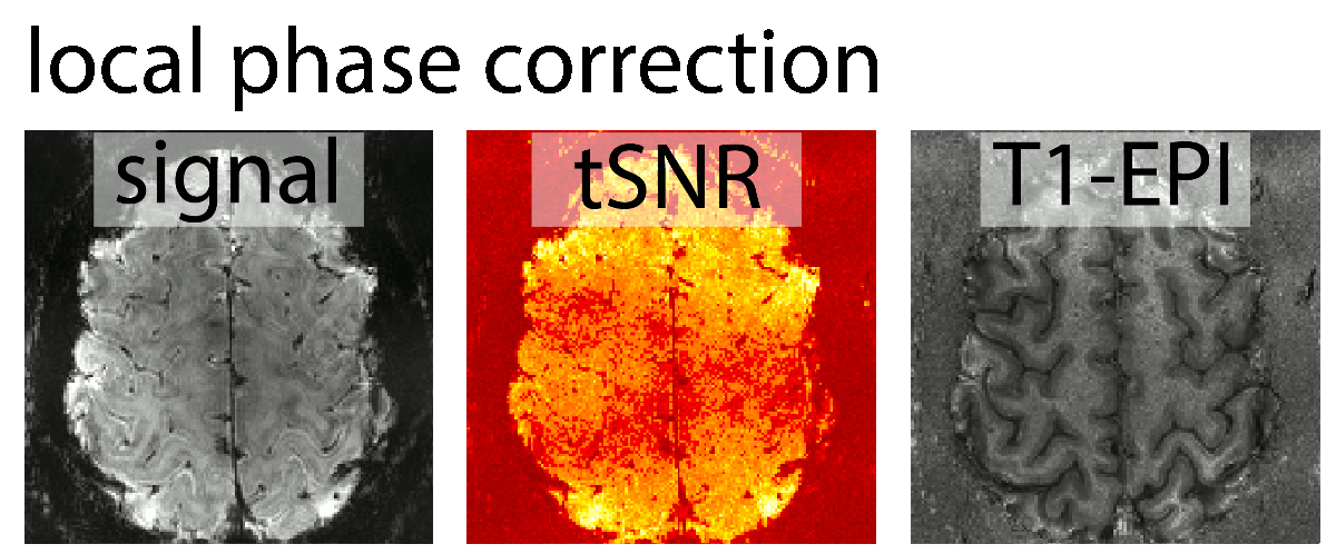
At high resolution EPI, the gradients are pushed to their limits and the ramp sampling ratio is particularly large. This means that the ghosting is increased and the Nyquist ghost correction is getting more important. In this post, I describe how to change the Nyquist ghost correction algorithm.
The high ramp sampling ratio in high-resolution EPI results in larger ghosts. Changing the correction algorithm from “normal” to “local” can help a lot.
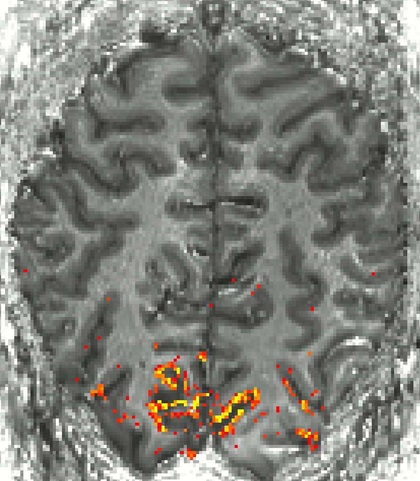
Activation maps for BOLD and VASO. At about 0.8 mm resolution, one starts to see that VASO is less sensitive to large draining veins.
With respect to high-resolution VASO application, visual cortex is very unique. I found it to be a challenging area. However, because of its high demand, I have been working on is with multiple collaborators. The most important pitfalls of SS-SI VASO in visual cortex that I came across in these collaborations are discussed below.
The take home message tat I learned from manny experiments is:
Use axial slices with the phase encoding direction A>>P.
Watch out for negative voxels.
Invest a lot of effort in optimizing GRAPPA parameters, its worth it.
This blog post discusses the resolution loss when applying partial-Fourier imaging in GE-EPI in the presence of strong T2*-decay.
I found that that when I was aiming for high-resolutions, it is beneficial to refrain from the application of partial Fourier (PF) imaging as much as possible. For the long readout durations at high-resolutions and the fast T2/T2*-decay at high field strengths results in even stronger blurring of partial-Fourier.
In this post I want to describe the guidelines that helped me to find the right spot of primary motor cortex (M1) that has a double-layer pattern during a conventional finger tapping task.
The motor cortex is an excellent model system to debug-layer fMRI methodology for multiple reasons:
It has a consistent folding pattern across people.
Its folding pattern is convoluted across one axis only. Hence, it is possible to use thicker slices with higher in-plane resolution.
With 4mm, its is the thickest part of the cortex compared to all other areas. Hence, layer analysis can be done even with 1.2 mm voxels.
It has an expected double layer structure, with two separate peaks. The separability of the peaks can be used as a measure of functional specificity.
It is very close to the RF-receive coils and has high tSNR.
It is easy to shim.
One tricky part, however, is to find the right location of the double layer feature.
I got interested in gradient temperature because of the weird effect that tSNR seemed to increase over time.
participant 1, tSNR maps refer to a 30 min fMRI run
Upon posing this effect on Twitter, PracitalfMRI and Ben Poser suggested that it might be due to gradient temperature. So I learned how to track it with as described below.
Almost every modern fMRI protocol (at SIEMENS scanners) uses GRAPPA. However, only very few people pay a lot of attention on optimal usage of the GRAPPA auto-callibration data. I realized the importance of optimizing GRAPPA parameters when doing high-resolution EPI. At high resolutions, GRAPPA-related noise can become an increasingly important limitation. This is especially true with the low bandwidth that the body gradient coils force us to use.
In this blog-post I will explain how the GRAPPA kernel-size affects the fMRI data quality, how you can change it, how you can find out which kernel-size was used, and I will descrive simple software tools to identify regions that might benefit from adaptations of the GRAPPA-kernel size.


